Module 2 - Nerves, Vessels and Lymphatic Drainage: Page 2 of 7
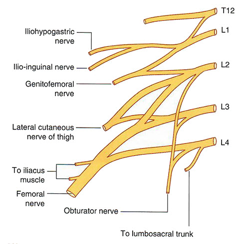
Add the obturator nerve. Rotate the 3D model to follow the course of the obturator nerve as it exits the pelvis through the obturator canal, a small opening in the obturator foramen. The obturator nerve supplies the medial compartment of the thigh and has key relationships within the pelvic cavity.
Add the femoral nerve. Visualize its path along the lateral pelvic wall before it passes beneath the inguinal ligament to enter the anterior thigh.
Pay close attention to the following nerves derived from the lumbar and sacral plexuses: obturator nerve, femoral nerve, genitofemoral nerve, pudendal nerve, coccygeal nerves, and pelvic splanchnic nerves. Note that both the femoral and obturator nerves originate from spinal nerves L2–L4, despite having very different destinations and functions.