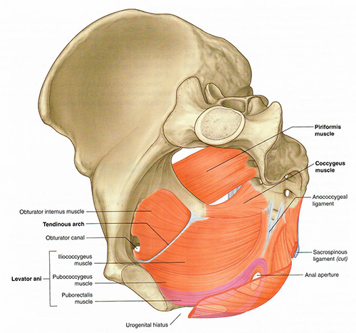
Add the coccygeus muscle. This triangular muscles extends from the ischial spine to the sacrum and coccyx, completing the posterior portion of the pelvic diaphragm.
Add the internal anal sphincter. This smooth muscle surrounds the anal canal and plays a key role in involuntary control of defecation.
Steps 14, 15 and 16 complete the Pelvic Floor.
Rotate the pelvis to see where the levator ani joins the obturator internus. This site is known as the tendinous arch of the levator ani, a thickened fascia of the obturator internus. It delineates the lateral boundary of the pelvic floor.
Rotate the pelvis to view the perineum.
Tap on image to enlarge
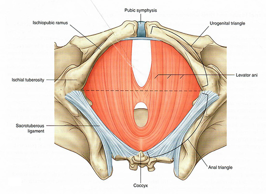
Draw an imaginary line between the ischial tuberosities. This line divides the perineum into the anal triangle (posteriorly) and the urogenital triangle (anteriorly). It thus separates the posterior muscular pelvic diaphragm, which controls defecation, from the anterior urogenital triangle which contains the vaginal and the urethral openings.
What are the bony and soft tissue landmarks that delineate the urogenital triangle?
Anteriorly, the pubic symphysis.
Laterally, the ischiopubic rami. The base of the triangle is the imaginary line connecting the two ischial tuberosities.
Posteriorly, the imaginary line connecting the ischial tuberosities.