Female Pelvis and Genitalia: Module 1 - Page 6 of 7
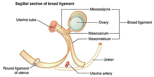
In this section, you will identify components of the broad ligament, a peritoneal fold that helps suspend the uterus, ovaries, and uterine tubes. These structures are not visible in the 3D model but using the anatomical illustration provided identify th efollowing structures that form it:
Tap on image to enlarge
Examine the mesovarium, the portion of the broad ligament that suspends the ovary and conveys the ovarian vessels.
View the mesosalpinx, the superior portion of the broad ligament that encloses the uterine (fallopian) tubes.
Identify the mesometrium, the largest portion of the broad ligament that supports the uterus itself.
Add the urinary bladder and highlight the ureters.
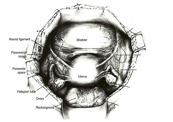
Put all of this information into a surgical context:
This is a view of the pelvis commonly encountered by OB/GYN surgeons during pelvic surgeries. Understanding the spatial relationships and peritoneal reflections is critical to avoid damaging important structures like the ureters and uterine arteries.
Rotate the 3D model to match the surgical view of the pelvis in the illustration. Take a moment to orient yourself before you proceed.
Identify the following pelvic structures using the surgical illustration:
Using your imagination, mentally reconstruct the three major subdivisions of the broad ligament: