END 1 Lab 2: Female Pelvis and Genitalia: Module 2
OBJECTIVES:END1.3.1 Identify the extent of the peritoneal cavity, its folds, its reflections, and its pouches and spaces in the female pelvis and their relationship to the pelvic contents. END1.3.2 Distinguish between the ligaments which are formed by folds of peritoneum, ligaments formed by the gubernaculums, and ligaments which are formed by condensations of visceral pelvic fascia in terms of their function and location. END1.3.3 Identify the pelvic viscera, their normal positions and anatomical relationships. END1.3.4 Trace the course of the round ligament of the ovary as it passes through the inguinal canal to the labia majora. |
Female Pelvis and Genitalia: Page 1 of 7
Although the membrane covering the urogenital triangle is not visible in this model, its associated structures can be identified. These components include several included in the next steps.
 |
| Tap on image to enlarge |
| Begin where we left off in Lab 1. | |
| Add the superficial transverse perineal muscle. | |
| Add the vaginal orifice. |
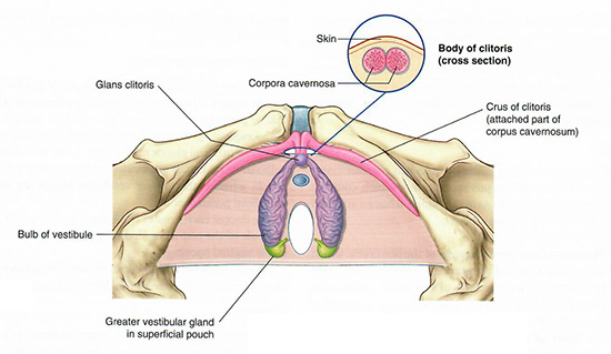
The erectile structures of the urogenital triangle are positioned on top of this membrane, as depicted in the illustration.