
Neurology Lab 2 - Module 1 - Vertebral Column: Page 5 of 5
Look at the accompanying diagram and answer the following questions:
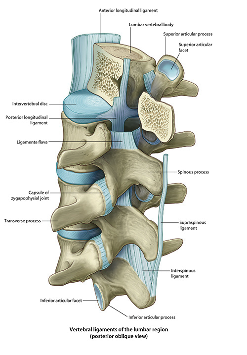
Examine the anterior longitudinal ligament (ALL) and posterior longitudinal ligament (PLL). Given that ligaments tend to stabilize joints, guide normal joint motion, and limit excessive or abnormal movements, consider the anatomical positions of the ALL and PLL - what would you predict they each do? |
|
|
The anterior longitudinal ligament extends along the anterior surface of the vertebral bodies and IV discs, from the occiput to the sacrum. It resists hyperextension and stabilizes the vertebral bodies. The posterior longitudinal ligament runs along the posterior surface of the vertebral bodies and intervertebral discs, within the vertebral canal, extending from C2 to the sacrum. The PLL prevents hyperflexion and limits posterior protrusion of the IV discs into the vertebral canal. |
|
In what direction do intervertebral discs most commonly herniate, and which anatomical features explain this pattern? |
|
|
IV discs most commonly herniate posterolaterally. The posterior longitudinal ligament runs along the posterior surfaces of vertebral bodies but is narrower than the anterior longitudinal ligament, leaving the posterolateral disc margins less supported. Moreover, the nucleus pulposus lies slightly posterior to the disc's center, making this region structurally more vulnerable to herniation. |
|