Anatomy Explorer Lab - Module 4 - Internal Brain: White Matter Page 1 of 1
Goals
Continue exploring the brain and remember to
click on the hyperlinks for bookmarked views, and follow the instructions in the VHD below.
Tap on image to enlarge
White Matter. Observe all of the highlighted white matter structures on the 3D model and in the cross-sections. We will explore specific structures in detail below.
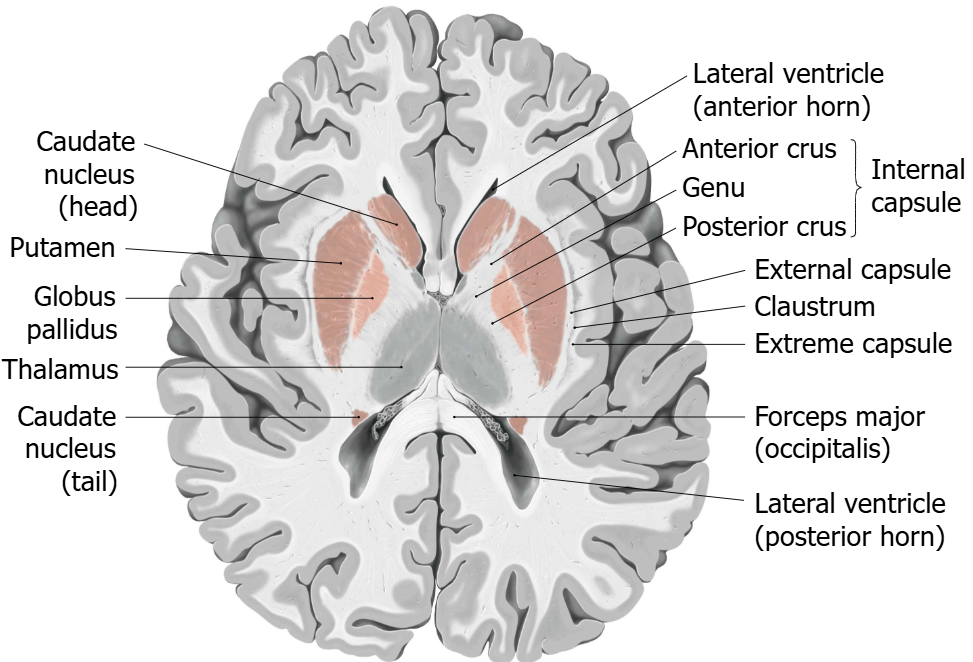
Internal Capsule. The internal capsule is the white matter tracts that run through the grey matter between the caudate and putamen causing a "striped" appearance. Hence the name striatum.
Broadly, it contains both ascending and descending tracts, running from the cerebral cortex to the spinal cord (or spinal cord to cerebral cortex).
Since this white matter tract plays a major role in the brain-body connection, lesions here can result in paralysis, weakness, and/or somatosensory loss on the contralateral side of the body.
Internal Capsule. Identify and highlight the internal capsule on the left and right sides of the brain.
Rotate the model and describe the relationship of the internal capsule to the basal ganglia.
Orient yourself to ventral, dorsal, rostral, and caudal.
Cross-Sections. Overlay the coronal, axial (transverse), and sagittal slices individually as you did in the above sections.
Use the 3D model to observe the internal capsule & its relationship to the MRI slices.
Observe the highlighted internal capsule in the cross-sections panel as you scroll through the MRI.
Corpus Callosum. The corpus callosum is a large bundle of white matter tracts that connect the left and right cerebral hemispheres and facilitates communication between the two sides of the brain.
In epilepsy, electrical seizures can spread from one side of the brain to the other. As a last resort, the corpus callosum can be surgically severed. Deficits that result from this surgery are explained by the sidedness of the human brain. For example, the left hemisphere (in most individuals) controls speech. If a patient sees an object only in the left half of their visual field (which is processed in the right occipital lobe), they cannot verbalize what they are looking at after this surgery because that information cannot reach the language areas in the left hemisphere.
Highlight the Corpus Callosum. Use the highlight tool to identify and observe the corpus callosum in the 3D model.
Rotate the model and describe the relationship of the corpus callosum to the other structures (thalamus, ventricular system, etc).
Orient yourself to ventral, dorsal, rostral, and caudal.
Anterior Commissure. The anterior commissure is a bundle of white matter tracts that connects the left and right temporal lobes, including the amygdala. Its function is to facilitate communication between the two sides of the brain in the temporal lobe region. It also serves to connect the olfactory tracts across the midline.
Highlight the Anterior Commissure. Use the highlight tool to identify and observe anterior commissure in the 3D model. Zoom in and out of the cross-sections to appreciate this bilateral structure in all three image planes.
Rotate the model and describe the relationship of the anterior commissure to the other structures (thalamus, hippocampus, etc).
Orient yourself to ventral, dorsal, rostral, and caudal.
Overlay the coronal, axial (transverse), and sagittal slices individually as you did in the above sections and scroll through the slices with the anterior commissure highlighted.
Fornix. The fornix is part of the limbic system. It consists of a bundle of white matter tracts that connect the hippocampus to the mammillary bodies. Note that it connects rostral-ventral structures, not left and right structures (in other words, it's not a commissure!).
Explore the Fornix.Rotate the model and describe the relationship of the fornix to the other structures.
Orient yourself to ventral, dorsal, rostral, and caudal.
Use the dissect tool to remove other structures to better view the fornix.
Note that the fornix forms the floor of the lateral ventricles while the corpus callosum forms the roof. Use the highlight tool to highlight these structures on the 3D model.
Now highlight the hypothalamus and the hippocampus on one or both sides to see how the fornix connects these structures.
Overlay the coronal, axial (transverse), and sagittal slices individually as you did in the above sections and scroll through the slices with the anterior commissure highlighted.
Coronal Section. Overlay the coronal stack individually as you did in the above sections and scroll through the slices with the fornix highlighted.
Axial Section. Overlay the axial stack individually as you did in the above sections and scroll through the slices with the fornix highlighted.
Coronal Section. Overlay the sagittal stack individually as you did in the above sections and scroll through the slices with the fornix highlighted.
Virtual Brain. CONGRATULATIONS!!! YOU'VE JUST BUILT A VIRTUAL BRAIN!
Review. View this model to test your knowledge! See if you can identify each colored structure (you can use the highlight tool to check your answers).
Also, scroll through the MRI cross-sections and see if you can identify each colored structure on both sides of the brain.