Anatomy Explorer Lab - Module 3 - Internal Brain: Gray Matter Page 2 of 3
Goals
Continue exploring the brain and remember to
click on the hyperlinks for bookmarked views, and follow the instructions in the VHD below.
Tap on image to enlarge
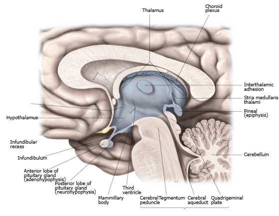
Thalamus. The thalamus relays sensory, motor, and limbic information. There are two thalami in the brain, one on each side.
Third Ventricle.
The two thalami sandwich the 3rd ventricle and are connected by a flattened band of tissue called the interthalamic adhesion.
Use the highlight tool on the coronal cross-section to identify and highlight the left, then right thalamus. Note the apparent "hole" in the 3rd ventricle marks the location of the interthalamic adhesion).
Each thalamus deals with contralateral body and ipsilateral cortex. There are many subnuclei in the thalamus.
The thalamus also plays a role in sleep, wakefulness, and consciousness. Damage or degeneration of the thalamus can result in coma or insomnia, respectively.
Rotate the thalamus model and describe its relationship to the caudate, putamen, globus pallidus, and ventricles.
Orient yourself to ventral, dorsal, rostral, and caudal. If needed, use the dissect tool to remove the caudate, putamen, and globus pallidus to better view the thalamus. (Tip: Use Undo/Redo under the Edit menu to toggle structures on/off with the dissect tool).
MRI Cross-Sections. Overlay the coronal slice, axial (transverse) slice, and sagittal slices individually.
Use the 3D model to observe the thalamus and its relationship to the MRI slices.
Observe the highlighted thalamus in the cross-sections panel as you scroll through the MRI. While viewing a coronal slice of the thalamus, the hippocampus will also be visible.
Hypothalamus. The hypothalamus maintains homeostasis by controlling autonomic output and pituitary hormones.
It regulates appetite, thirst, temperature/sweating, circadian rhythms, and reproductive/maternal behaviors - your “primal switchboard.”
Injury can cause pituitary hormone deficiencies (treat with hormone replacement) and Horner syndrome from sympathetic disruption: ipsilateral ptosis, miosis, anhidrosis.
Reset the Hypothalamus.
Use the highlight tool to identify and highlight the hypothalamus on the 3D model.
Rotate the model and describe the relationships of the hypothalamus to other structures, especially the 3rd ventricle and the thalamus.
Orient yourself to ventral, dorsal, rostral, and caudal.
Use the dissect tool for a better view (Tip: Use the Undo/Redo option when needed!).
Coronal MRI Cross Sections. Overlay the coronal slices.
Identify and highlight the hypothalamus on the cross-section images. Use the 3D model to observe the hypothalamus and its relationship as you scroll through the MRI slices.
While viewing a coronal slice of the hypothalamus, the amygdala will also be visible. Highlight the amygdala and scroll through the stack.
Axial MRI Cross Sections. Identify and highlight the hypothalamus on the cross-sections. Observe its relationships as you scroll through the stack.
Sagittal MRI Cross Sections. Identify and highlight the hypothalamus on the cross-sections. Observe its relationships as you scroll through the stack.
A patient has a lesion involving the hypothalamus. What two major downstream clinical consequences might result?
Damage to the hypothalamus can result in a malfunctioning pituitary gland, disrupting many basic bodily functions, which can be treated via hormone therapy. Damage to the hypothalamus can also result in Horner's syndrome, a disorder of the autonomic nervous system (which can have many other causes as well). The classic symptoms are ptosis, miosis, and anhidrosis on one side of the face.