Anatomy Explorer Lab - Module 3 - Internal Brain: Gray Matter Page 1 of 3
Goals
Continue exploring the brain and remember to
click on the hyperlinks for bookmarked views, and follow the instructions in the VHD below.
Tap on image to enlarge
Gray matter. Observe all of the highlighted grey matter structures on the 3D model and in the cross-sections. We will explore specific structures in detail below.
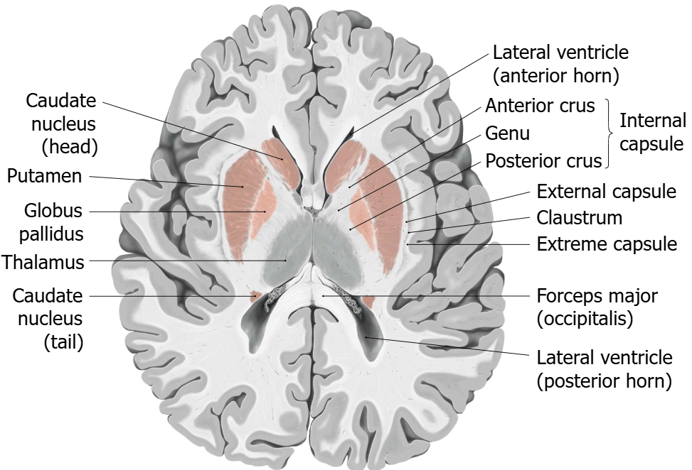
Basal Ganglia (Nuclei).
The basal ganglia are important for initiating motor movements. They include the caudate, putamen, globus pallidus, substantia nigra, and subthalamic nucleus.
In the VHD, only the caudate, putamen, and globus pallidus are clearly visible. This lab focuses on these structures which comprise the telencephalic portion of the basal ganglia.
The basal ganglia are associated with disordered movement conditions like Parkinson and Huntington Diseases.
Left Basal Ganglia. Highlight the caudate, putamen, and globus pallidus on the left side of the brain. Do so with the highlight tool or hover over structures with the dissect tool.
Orient yourself to ventral, dorsal, rostral, and caudal. Rotate the model and explore the relationships of these structures to each other and the ventricles.
Sagittal Cross-Section. Explore the cross section overlain on the 3D model. Identify the highlighted basal ganglia structures in the image.
Rotate and appreciate the 3D relationships to the 2D slices.
Use the highlight tool on the sagittal cross-sections to add the corresponding structures on the right side of the model.
MRI Cross-Sections. Overlay the coronal, sagittal, and axial cross-sections individually over the 3D model one at a time.
Observe the basal ganglia and their relationships. Scroll through the stacks and explore the highlighted structure within them.
Nucleus accumbens. Identify this structure where the caudate and putamen come together anteriorly.
Use the highlight tool on the axial slice to find and highlight the nucleus accumbens.
This structure is involved in many functions, its most notable relates to reward and addiction.
A patient with a lesion of the ventral basal ganglia develops decreased motivation and impaired reward-seeking behavior but has intact voluntary motor strength and coordination. Which major anatomical division of the basal ganglia is most likely involved?
The ventral striatum (including the nucleus accumbens)..