Anatomy Explorer Lab - Module 2 - Internal Brain: Ventricles - Page 1 of 1
Goals
Today, you'll get to explore the ins and outs of a brain from the comfort of the WFUSM virtual lab! The objectives of this lab are:
1. To identify the major internal brain structures in the three cardinal planes (coronal, sagittal, or axial) on a T1-weighted MRI.
2. To describe their spatial relationships (size, shape, orientation) and basic functions.
Click on the hyperlinks for bookmarked views, and follow the instructions in the VHD below.
Tap on image to enlarge
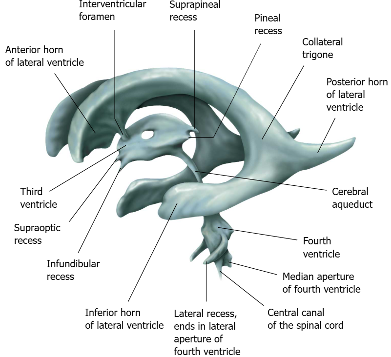
Start by examining the ventricles.
There are four ventricles in the brain: two lateral ventricles, a third ventricle, and a fourth ventricle.
The main function of the ventricles is to store and produce cerebrospinal fluid (CSF) via the choroid plexus. CSF acts as a shock absorber, protecting the brain from injury. Additionally, CSF "lavages" or washes the brain parenchyma removing waste around the clock but especially when you sleep.
Rotate the 3D structure and orient yourself to the caudal, rostral, ventral, and dorsal directionality of the ventricles.
Identify the following structures:
Now, visually trace the pathway of 1 drop of CSF through the ventricular system, starting at the choroid plexus in the lateral ventricle.
Explore the coronal cross-section.
Use the highlight tool to identify and highlight the ventricles in the coronal image. Which region(s) of the ventricular system is visible in this image?
Use the 3D mode to observe the relationships of the ventricles as you scroll through the coronal MRI slice. What portion(s) of the ventricular system is visible in the coronal section?
Using anatomical terms of directionality, describe the direction the coronal plane moves through the head.
Remember, if you want to add the section to the 3D model, click the filter button and select the coronal axis.
Explore the sagittal cross-section and remove the axial section.
Identify and highlight the ventricles in the sagittal image. Which region(s) of the ventricular system is visible in the image?
Use the 3D mode to observe the ventricles and their relationships to the sagittal MRI slice.
Observe the highlighted ventricles in the sagittal MRI as you scroll through the cross-sections.
Using anatomical terms of directionality, describe the direction the axial plane moves through the head.
Explore the axial (transverse) cross-section and remove the coronal section.
Identify and highlight the ventricles in the sagittal image. Which region(s) of the ventricular system is visible in the image?
Use the 3D mode to observe the ventricles and their relationships to the sagittal MRI slice.Observe the highlighted ventricles in the sagittal MRI as you scroll through the cross-sections.
Using anatomical terms of directionality, describe the direction the sagittal plane moves through the head.
Explore all cross-sections together. Remember, you can focus on one cross-section or open all three by tapping the left panel when not in dissection mode.
When CSF flow is obstructed or (less commonly) when CSF is overproduced, hydrocephalus develops, leading to ventricular enlargement and increased intracranial pressure. How is hydrocephalus treated?
Hydrocephalus most commonly results from obstruction of CSF flow (noncommunicating hydrocephalus). Treatment is CSF diversion with a VP shunt. Endoscopic third ventriculostomy is an alternative in selected obstructive cases. CSF overproduction is rare (e.g., choroid plexus papilloma).