Lab 8 - Module 3 - Anatomy of the Dorsum of the Foot: Page 2 of 4
×
Arteries on the Dorsum of the Foot
Tap on image to enlarge
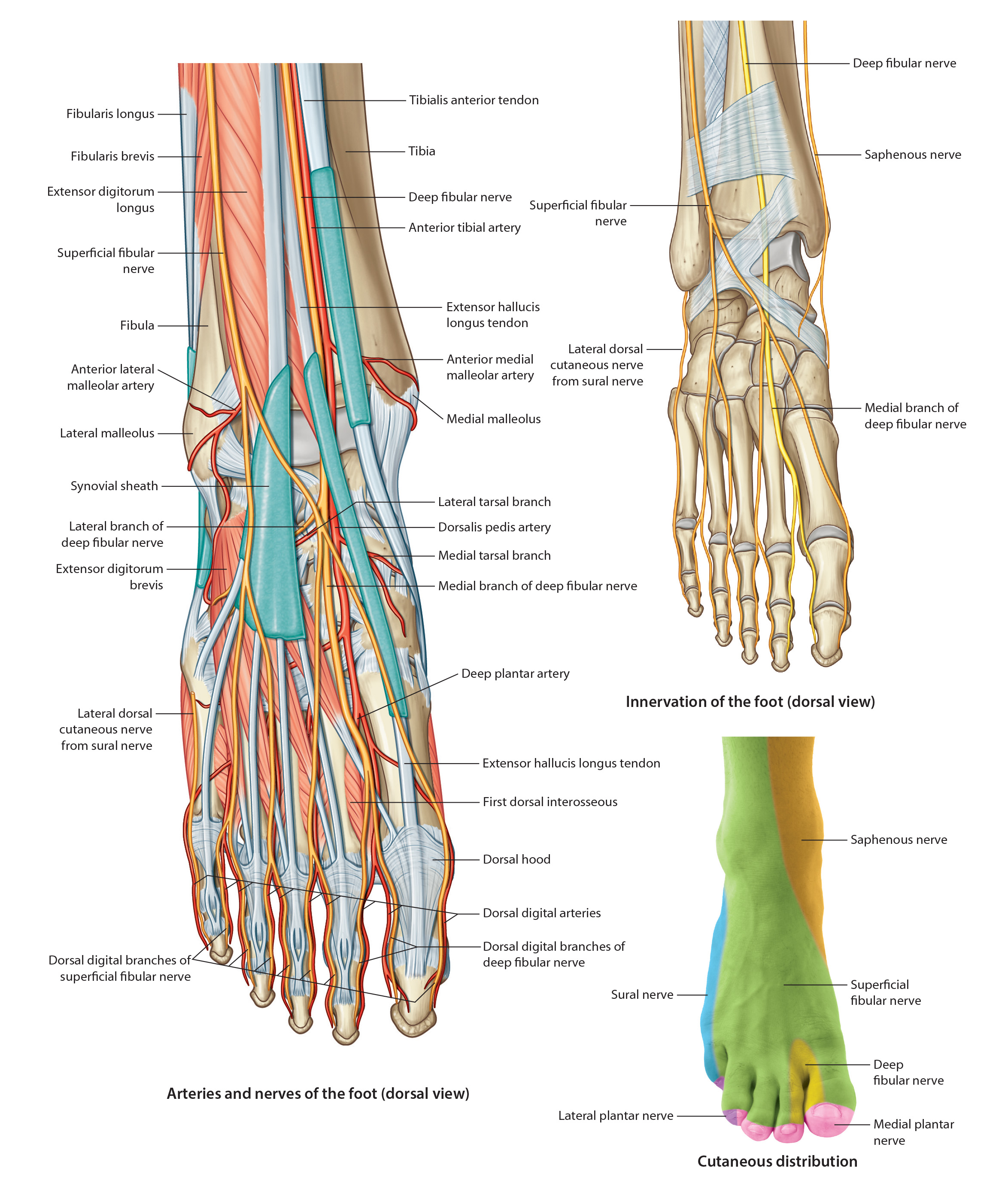
The arteries of the dorsum of the foot are small and not identified on the 3D cadaver. See diagram for location of each artery.
• The anterior tibial artery crosses the dorsal surface of the ankle joint and is now called the dorsalis pedis artery. The dorsal pedis artery travels distal, just lateral to the extensor hallucis longus tendon, to the space between the first and second metatarsal bases. There it branches into a deep plantar artery that dives deep into the space to the plantar aspect of the foot (to anastomose with the lateral plantar artery) and an arcute artery that travels lateral across the dorsum of the bases of the lateral four metatarsals. It may anastomose with the lateral tarsal artery.
• The lateral tarsal artery branches off of the dorsal pedis artery and runs lateral and inferior, deep to the EDB. It anastomoses with the arcuate artery (sometimes) as well as the anterior lateral malleolar artery. A perforating branch of the fibular artery emerges between the distal tibia and fibula to join this anastomosis.
• From the arcuate artery arises the second, third, and fourth dorsal metatarsal arteries. The first dorsal metatarsal artery arises from the division of the dorsal pedis artery. Each metatarsal artery divides into two dorsal digital arteries.