Albany Medical College Virtual Anatomy Lab

Lab 8 - Module 2 - Anatomy of the Anterior and Lateral Compartments: Page 1 of 3

READINGS:

Gray's Anatomy for Students (Fourth Edition): Pages: 624-627


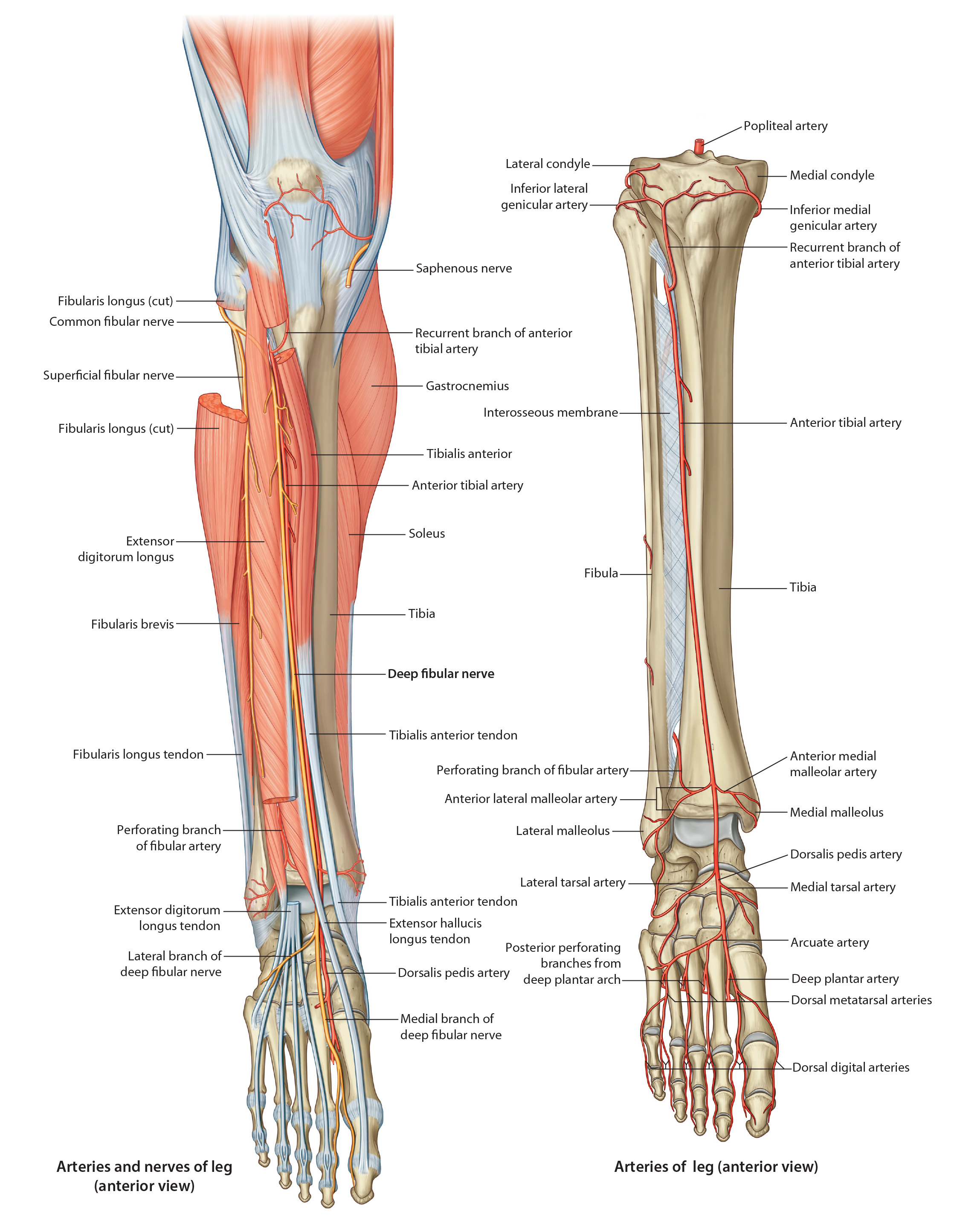
Nerves and Vessels of the Anterior Compartment

naantlat
naantlat
Tap on image to enlarge
Anterior compartment is located anterior to the interosseous membrane between the lateral surface of the tibia and anterior crural intermuscular septum.

It contains the dorsiflexors of the ankle and extensors of the toes.


Begin with an anterior view of the Anterior Leg.
Add the Anterior Tibial artery which comes off the common tibial artery from the popliteal artery. The anterior tibal artery passes between two heads of the posterior tibialis muscule on the interosseus membrane. The artery pierces the membrane then lies on its anterior surface, deep to the Extensor Digitorum Longus. The anterior tibial artery terminates at the inferior extensor retenaculum as it becomes the dorsalis pedis artery.
Add the Anterior Tibial veins which is a paired system parelling the anterior tibial artery. .
Add the Common Fibular Nerve (Peroneal) which comes around the head of the fibula to enter the lateral compartment of the leg. While in the fibularis (peroneus) longus muscle, the common fibular nerve will split into the superficial and deep branches.
Add the Deep Fibular Nerve (eroneal) which innervates all the muscles of the anterior compartment. • The deep branch travels out of the lateral compartment and enters the EDL in the anterior compartment. It descends through the anterior compartment on the interosseous membrane and will supply all four anterior compartment muscles and the EDB and EHL on the dorsum of the foot. The deep peroneal nerve's sensory portion exists in the first web space of the dorsum of the foot.
The Superficial Fibular Nerve (Peroneal) . (NOT IDENTIFIED IN THIS CADAVER)