Lab 8 - Module 1 - Anatomy of the Knee: Page 4 of 10
Osteology of the Knee
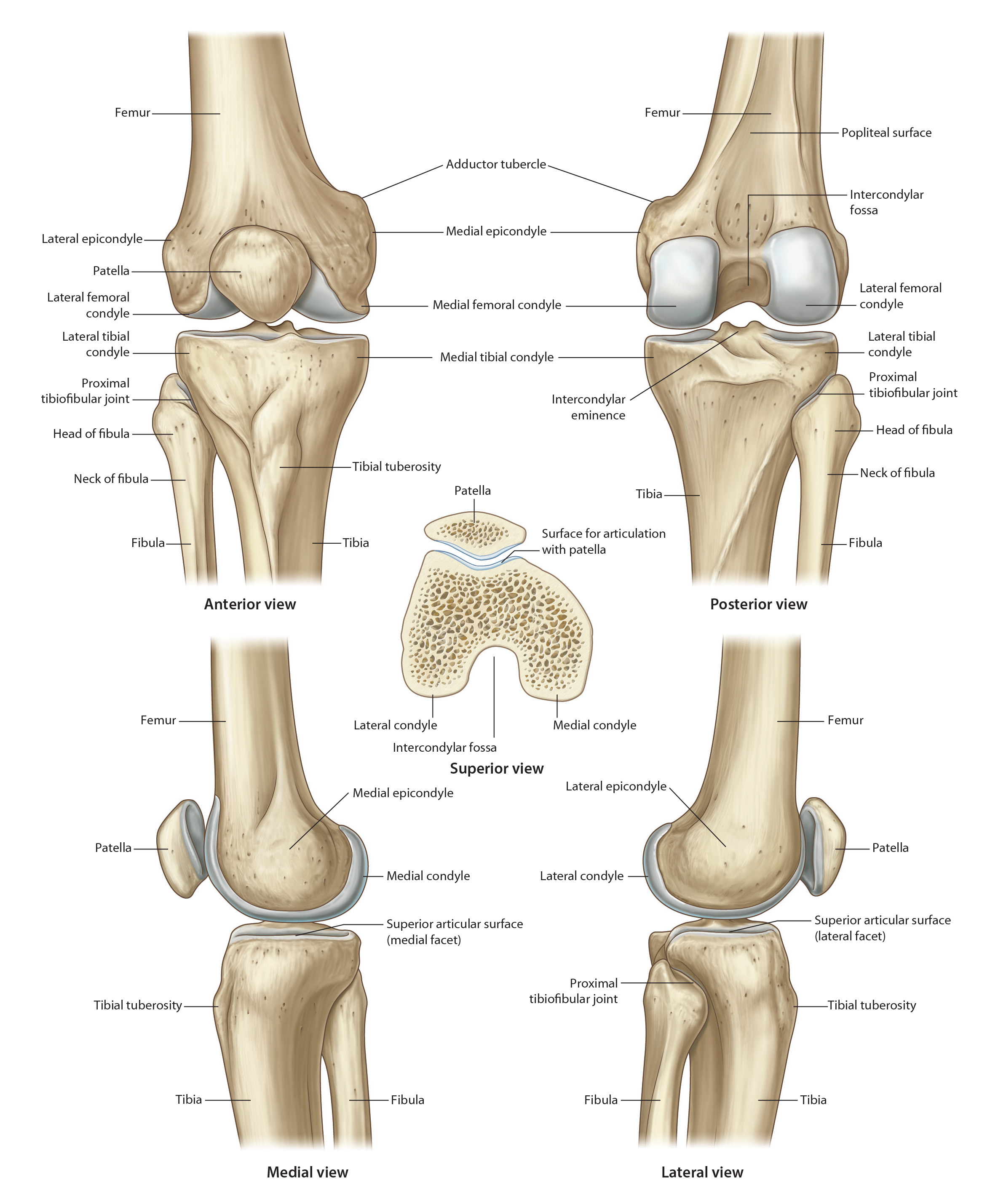
Tap on image to enlarge
Add the fibula. (Rotate the femur to visualize the following land marks:)
The fibula is the lateral and more slender of the two bones of the leg. Its proximal expanded end is the head. Its distal expanded and somewhat flattened end is termed the lateral malleolus and is part of the ankle (talocrural) joint. The head and lateral malleolus of the fibula are both readily palpable. Between the proximal and distal ends of the fibula is the elongated and slender shaft of the fibula. The interosseous border on the antero-medial aspect of the fibular shaft is joined to the interosseous border on the antero-lateral aspect of the tibial shaft by the interosseous membrane whose fibers are oriented in an infero-lateral direction.
The junction between the head of the fibula and the shaft is termed the fibular neck.
The head of the fibula is the expanded proximal end. It has the shape of an irregular cuboid. Its anterior, lateral and posterior aspects are palpable. Projecting proximally from its postero-lateral aspect is a conical process called the styloid process. Attached to the styloid process is the arcuate ligament.
The superomedial aspect of the head of the fibula displays a smooth facet. This articulates with a corresponding facet on the inferior aspect of the posterolateral part of the lateral tibial condyle to form the superior tibiofibular articulation, a synovial articulation. Rarely the synovial cavity of the superior tibio-fibular joint may communicate with that of the knee joint.
The lateral surface of the head of the fibula receives the attachments of the lower end of the fibular (lateral) collateral ligament and the tendon of biceps femoris, the attachment of biceps femoris being in front of the attachment of the fibular collateral ligament. Posterior to the attachment of the fibular collateral ligament, this surface receives the attachment of the menisco-fibular ligament when this ligament is present. Infero-medial to the styloid process, the posterior surface of the fibular head often presents a groove caused by the popliteus tendon on its way to the lateral femoral condyle. An important relationship to the postero-lateral aspect of the fibular head is the common peroneal nerve.
The styloid process of the fibula is a conical, upward projection of bone on the postero-lateral aspect of the fibular head. It is sometimes palpable. The arcuate ligament is attached to it.
The neck of the fibula is the constriction which marks the junction between the head and shaft of fibula. It is thinly wrapped in muscle; peroneus longus laterally, extensor digitorum longus anteriorly and soleus posteriorly. A very important and close topographical relation is the common fibular nerve as it winds around the lateral aspect of the fibular neck.
The shaft of the fibula presents a fairly generous surface on its lateral aspect called the peroneal surface. Peroneus longus arises from the upper two-thirds while peroneus brevis arises from the lower two-thirds, of this surface. The peroneal surface of the fibula is limited by anterior and posterior borders. Medial to the anterior border of the fibula and very close to it is the interosseous border of the fibula. Between the anterior border and interosseous border, is the rather narrow extensor surface of the fibula. Extensor digitorum longus arises from the upper three-fourths of this surface while peroneus tertius arises from the lower quarter of this surface.
Between the interosseous border and the posterior border on the medial aspect of the fibular shaft is the posterior or flexor surface of the fibula. This surface gives attachment to flexor hallucis longus and tibialis posterior. A strip of bone on the upper part of this surface gives attachment to soleus.
The medial aspect of the lower end of the fibular shaft presents a roughened area. Joining this area with a corresponding roughened area on the lateral aspect of the lower end of the tibia is a strong ligament called the interosseous tibio-fibular ligament.
The lower end of the fibula, also called the lateral malleolus, presents a subcutaneous and easily palpable lateral surface. The medial surface of the lower end of the fibula has a smooth triangular facet which articulates with a reciprocal facet on the lateral aspect of the body of the talus, this being part of the talocrural articulation. Posterior to the triangular articular facet is a fossa termed the malleolar fossa. This gives attachment to ligaments which contribute to the stability of the talocrural articulation.
Superior tibio-fibular joint:
The superior tibio-fibular joint may be dislocated traumatically and in this case, the peroneal nerve may be vulnerable. Clicking and recurrent subluxation of this joint may occur following the initial trauma. The resultant pain may be helped by local injection of anesthetic plus hydrocortisone.