Lab 8 - Module 1 - Anatomy of the Knee: Page 5 of 10
×
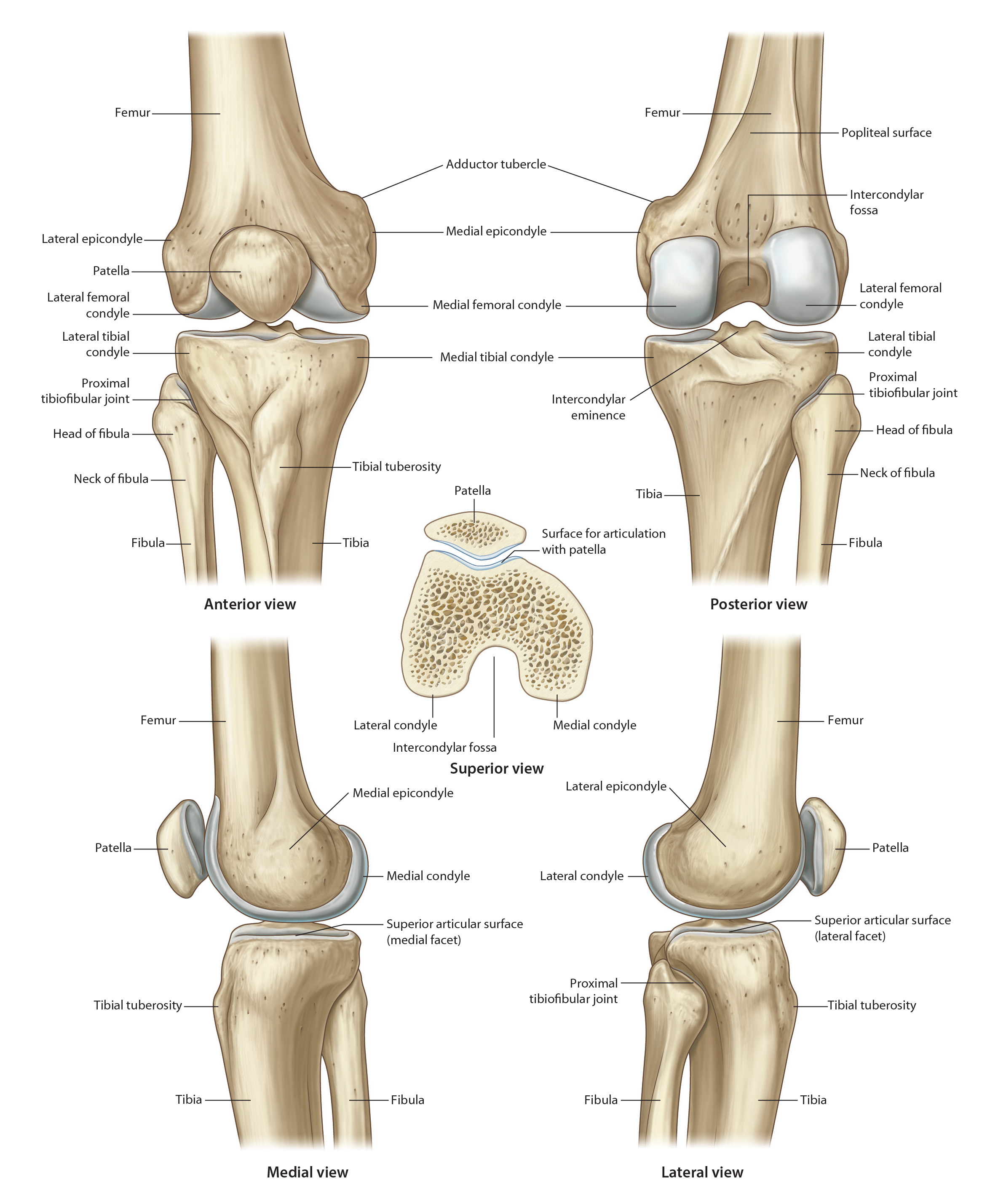
Osteology of the Knee
Tap on image to enlarge
Add the patella. (Rotate the femur to visualize the following land marks:)
The patella, the largest sesamoid bone in the body, is embedded in the tendon of quadriceps femoris, and is located anterior to the tibio-femoral articulation. Its outline is somewhat in the shape of an inverted triangle with the apex of the bone directed inferiorly. Thus it possesses a base, lateral and medial borders and an apex. The base is a relatively expansive surface and receives the lower end of the quadriceps tendon.
The patella is part of the knee joint extensor mechanism.
The patella has an anterior surface which is gently convex forwards. It often shows a number of small perforations, (for nutrient vessels), and a few ridges, but is otherwise featureless.
The posterior surface of the patella carries a large, smooth, undulant and quadrangular articular area. This facet occupies the upper four-fifths or so of the posterior surface of the patella, and is covered by a continuous plate of articular hyaline cartilage. A vertical ridge on this articular surface divides the area into lateral and medial facets, the lateral facet usually being larger and significantly more concave. These two facets of the patellar articular surface are apposed to the corresponding femoral condyles, while the vertical ridge separating them faces the groove on the anterior aspect of the lower end of femur between the two femoral condyles.
The medial patellar articular facet is further subdivided into two narrow vertical regions by means of a faint ridge, the medial of the two regions coming into contact with the medial femoral condyle in extreme flexion of the knee. There is also an “odd” facet that lies most medially. It articulates with the femur in deep flexion.
Attached to the apex of the patella and the adjoining part of the posterior patellar surface is the patellar ligament, the inferior continuation of the quadriceps tendon. Attached to the medial and lateral borders of the patella are, respectively, the medial and lateral patellar retinacula. Superficial to these retinacula, the medial and lateral patellar borders receive the attachments of the lowest fibers of vastus medialis and vastus lateralis respectively.