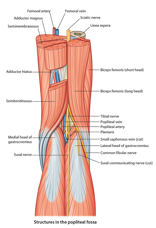
Add the Sciatic Nerve which ends at the superior angle of the popliteal fossa (usually but not always) by dividing into its terminal branches: tibial and common fibular (peroneal) nerves.
Add the Tibial Nerve which is the largest of the two terminal branches of the sciatic nerve. The motor portion of the tibial nerve supplies the muscles of the superficial and deep posterior compartments. iv. As the tibial nerve travels distal to the medial malleolus of the ankle (in the tarsal tunnel) it divides into the medial and lateral plantar nerves that provide sensory and motor to the plantar aspect of the foot.
Add the Sural Nerve. The medial sural nerve branches off of the tibial nerve and joins with a communicating branch from the common fibular nerve to form the sural nerve.
The medial sural nerve is cutaneous and supplies the skin of the proximal lateral calf.
The sural nerve provides sensation for the rest of the lateral calf to the lateral ankle and the base of the fifth metatarsal.
Add the Common Fibular (Peroneal) Nerve which is smaller of the two terminal branches of the sciatic nerve.
It provides a communicating sensory branch to the medial sural nerve to form the sural nerve.
Additionally, it has a lateral sural nerve that supply sensory to a small area of the proximal lateral leg.
As it leaves the popliteal fossa it passes superficial to the lateral head of the gastrocnemius muscle and superficial to the neck of the fibula. As a result the common fibular nerve is the most commonly injured nerve in the lower limb. Injury to this nerve results in the classic clinical presentation of foot drop.
As it spirals around the neck of the anterior fibula, it divides into a superficial fibular (peroneal) and deep fibular (peroneal) nerve.
Add the Superficial Fibular Nerve which supplies motor to the lateral compartment of the leg and sensory to the lateral leg and most of the dorsum of the foot. (NOT IDENTIFIED IN THIS CADAVER)
Add the Deep Fibular Nerve which supplies motor to the muscles of the anterior compartment of the leg, the two muscles that originate on the dorsum of the foot, and sensory to the first web space between the great toe and second toe.