Indentify the following:
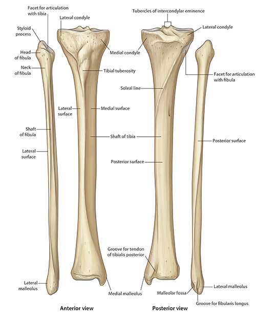
Tibial Plateau - The superior surface of the proximal end of the tibia is termed the tibial plateau.
Tibial Tuberosity - The anterior aspect of the upper end of the tibia, a short distance below the level of the plateau, is characterized by the presence of an irregular prominence, the tibial tuberosity. The upper half of the tibial tuberosity is smooth and is the site of attachment of the patellar ligament (patellar tendon). The lower half of the tibial tuberosity is rough and is overlain by the subcutaneous (superficial) infrapatellar bursa.
Shaft of the Tibia - The shaft of the tibia is approximately triangular in cross section, and may be described as possessing three borders - anterior, lateral and medial. The anterior border of the tibial shaft is colloquially referred to as the shin, and is readily palpable along its entire length. The lateral border of the tibial shaft gives attachment to the medial edge of the interosseous membrane and is therefore also referred to as the interosseous border.
The Medial Surface - The medial surface of the tibial shaft is almost entirely subcutaneous. The upper part of this surface receives the attachment of the pes anserinus (the partially conjoined tendons of insertion of sartorius, gracilis and semitendinosus). The upper part of the medial surface of the tibial shaft also receives the attachment of the lower end of the tibial collateral ligament, postero-inferior to the insertion of the pes anserinus.
The Lateral Surface - The lateral surface of the tibial shaft forms part of the floor of the anterior (extensor) compartment of the leg. The upper two-thirds of the lateral surface of the tibia gives attachment to tibialis anterior.
The Posterior Surface - The posterior surface of the tibial shaft displays, in its upper part, an oblique ridge which runs infero-medially from just below the lateral tibial condyle to the medial border of the tibial shaft. This ridge is called the soleal line and gives attachment to part of the long linear attachment of the soleus muscle.
The Triangular Area -The triangular area on the posterior surface of the tibial shaft proximal to the soleal line gives attachment to the popliteus muscle. Below the soleal line, the posterior surface of the tibia displays a vertical ridge which fades out distally. Tibialis posterior takes origin from the area lateral to the vertical ridge while flexor digitorum longus takes origin from the area medial to the ridge. The junction between the proximal three-fourths and distal fourth of the tibial shaft is the narrowest and weakest part of the tibial shaft. It is significant that beyond this junction, the tibial shaft has no muscular attachments on any of its surfaces. Consequently the periosteum of the distal tibia is less vascularized than in the proximal part of the bone. This has unfavorable implications for healing of fractures in this region. The medial aspect of the lower end of the tibia shows a prominent and thick process which projects distally. This is called the medial malleolus. The distal surface of the tibia is characterized by a smooth facet which is coextensive with a smooth area on the lateral surface of the medial malleolus. These surfaces articulate with the talus at the talocrural (ankle) joint. The lateral aspect of the distal part of the tibial shaft shows a prominent roughened area. This area gives attachment to the interosseous tibiofibular ligament, a very strong ligament which holds together the lower ends of the tibia and fibula.
The Lateral Condyle - The lateral condyle of the tibia is shorter from anterior to posterior than the medial tibial condyle. Since the lateral tibial articulation is smaller, the portion of the femoral condyles which articulates with the tibia is shorter from anterior to posterior on the lateral side of the joint.
The Medial Condyle - Understanding that the lateral side has less space available for articulation, we can understand that, as the joint moves from flexion into extension, the medial condyle of the femur can roll farther than the shorter lateral side. This is called the “Screw Home” Mechanism which will be discussed in detail below.