Lab 5 - Module 2 - Hip and Anterior Thigh - Muscles, Nerves and Vessels: Page 3 of 8
×
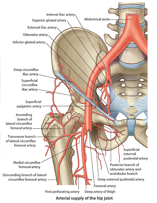
Branches of the Femoral Artery
Tap on image to enlarge
Add the Profunda Femoris. (NOT IDENTIFIED IN THIS CADAVER). The profunda femoris (deep femoral) artery is the largest and most important of the many branches of the femoral artery and is given off in the femoral triangle. It arises from the lateral aspect of femoral artery about 3.5cm below the inguinal ligament and is initially lateral to and then posterior to the femoral artery. Vascular surgeons and radiologists usually refer to that part of the femoral artery proximal to the origin of the profunda femoris artery as the ‘common femoral artery’, and the distal segment of the femoral artery as the ‘superficial femoral artery’.
Proximal to the origin of the profunda femoris artery, the femoral artery gives a variable number of relatively small, superficial branches. It gives rise to the superficial epigastric and superficial external iliac arteries just below the inguinal ligament, with the superficial external pudendal artery arising from the medial aspect of the femoral artery close by. The deep external pudendal artery also arises medially at a slightly lower level. The medial circumflex femoral artery usually arises from the arteria profunda femoris, but may arise from the femoral artery.
The femoral artery runs infero-medially, leaving the femoral triangle at its apex to enter the adductor canal in the medial part of the middle of the thigh. It is covered by muscle in the adductor (subsartorial) canal, with vastus medialis antero-laterally and sartorius antero-medially. In the distal part of the adductor canal the femoral artery gives off a branch called the ‘descending genicular artery’, which contributes to the anastomosis around the knee joint. The femoral artery then leaves the adductor canal through the adductor hiatus (a gap in the tendon of adductor magnus) to enter the popliteal fossa as the popliteal artery.
Add the Lateral Circumflex Femoral Artery which arises from the lateral side of the profunda femoris artery. It passes laterally behind sartorius and rectus femoris, and divides into ascending, transverse and descending branches.
Add the Ascending Branch of the Lateral Circumflex Femoral Artery which passes upwards to the lateral side of the hip, anastomosing with the superior gluteal and deep circumflex iliac arteries.
Add the Descending Branch of the Lateral Circumflex Femoral Artery runs downwards behind rectus femoris and along the anterior aspect of vastus lateralis, which it supplies. The branch descends to the knee, anastomosing with the lateral superior genicular artery.
A small transverse branch passes in front of vastus intermedius. It pierces vastus lateralis just below the greater trochanter to take part in the cruciate anastomosis with the medial circumflex femoral, inferior gluteal and first perforating arteries.
Add the Medial Circumflex Femoral Artery arises from the posterior medial aspect of the profunda femoris or femoral artery. It passes backwards between the psoas major and pectineus, and then between obturator externus and adductor brevis, reaching the upper border of adductor magnus.
It gives a branch to the hip joint and then divides into ascending and transverse branches. The ascending branch passes between obturator externus and quadratus femoris into the trochanteric fossa, to anastomose with the gluteal artery. The transverse branch passes between quadratus femoris and adductor magnus to join the cruciate anastomosis.
The Artery to the Head of the Femur is a branch of the obturator artery and traverses the ligament of the head. (NOT IDENTIFIED IN THIS CADAVER)
The Retinacular Arteries are the main blood supply to the hip joint. They are branches of the medial and lateral circumflex femoral arteries. The most abundant and important retinacular arteries arise from the Medial Circumflex Femoral artery. The retinacular arteries from the lateral circumflex femoral artery are smaller, fewer in number and have to traverse the thick iliofemoral ligament (Ligament of Bigelow). (NOT IDENTIFIED IN THIS CADAVER)
The Cruciate Anastomosis is a combination of the medial and lateral circumflex arteries, inferior gluteal artery, and the first perforating branch of the profunda femoris artery. They supply the thigh muscles and proximal end of the femur. (NOT IDENTIFIED IN THIS CADAVER)
How does the fracture, described in this case, impact the blood supply to the head/neck of the femur?
Compose an answer to help focus your study of this module.
How is this fracture likely going to be treated?
Compose an answer to help focus your study of this module.