
Lab 5 - Module 2 - Hip and Anterior Thigh - Muscles, Nerves and Vessels: Page 4 of 8
×
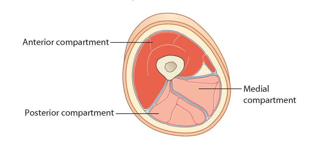
![Image]()
Anterior Compartment of the Thigh
 |
| Tap on image to enlarge |
| The Muscles of the Anterior Thigh – act as knee extensors and hip flexors. They are all innervated by the femoral nerve EXCEPT: psoas major (ventral rami of L1-L3) and Tensor Fasciae Lata (superior gluteal nerve). |
| Add the Psoas Major muscle. |
|
Origin - Sides of T12 – L5 vertebrae and the discs between them; transverse processes of all lumbar vertebrae Insertion - Lesser trochanter of femur as a conjoined tendon with iliacus Innervation - Ventral rami of L1, L2, L3 Action - Flex the thigh at the hip joint and aid in stabilizing the joint |
| Add the Iliacus muscle. |
|
Origin - Iliac crest, iliac fossa, ala of sacrum, and anterior sacroiliac ligaments Insertion - Con-joined with psoas major and inserts into lesser trochanter Innervation - Femoral Nerve Action - Flex the thigh at the hip joint and aid in stabilizing the joint |
Quadraceps Femoris Muscle Group
| Add the Vastus Medialis muscle. |
|
Origin - Intertrochanteric line and medial lip of linea aspera of femur Insertion - Distally, the muscle fibers become continuous with an aponeurosis, which joins the quadriceps tendon. A part of the vastus medialis aponeurosis is inserted directly into the medial edge of the patella. This is an important factor in maintaining patellar stability. A further expansion from the vastus medialis aponeurosis blends with the medial part of the capsule of the knee joint before attaching to the medial aspect of the medial tibial condyle. Innervation - Femoral Nerve (L2, L3, L4) Action - Vastus medialis, together with the other muscles that make up quadriceps femoris, extends the leg at the knee joint. It also aids in patella tracking and stability. |
| Add the Vastus Intermedius muscle. |
|
Origin - Vastus intermedius arises from the anterior and lateral surfaces of the proximal two-thirds of the femoral shaft. Insertion - Distally, the muscle becomes an aponeurosis, which in turn continues into the quadriceps femoris tendon. Innervation - Femoral Nerve (L2, L3, L4) Action - Vastus Intermedius, together with the other muscles that make up quadriceps femoris, extends the leg at the knee joint. |
| Add the Vastus Lateralis muscle. |
|
Origin - Greater trochanter and lateral lip of linea aspera of femur Insertion - Distally, the flattened tendon of vastus lateralis joins the quadriceps femoris tendon. A part of the tendon of vastus lateralis inserts directly into the upper and lateral borders of the patella. An expansion from the vastus lateralis tendon blends with the lateral aspect of the capsule of the knee joint and the iliotibial tract, before attaching to the lateral tibial condyle. Innervation - Femoral Nerve (L2, L3, L4) Action - Vastus lateralis, together with the other muscles that make up quadriceps femoris, extends the leg at the knee joint. |
| Add the Rectus Femoris muscle. |
|
Origin - AIIS and the ilium just superior to the acetabulum Insertion - Distally, a thick tendon develops from the muscle and joins the quadriceps femoris tendon, through which it attaches to the superior border of the patella, which then proceeds to the patella tendon ultimately inserting into the tibial tuberosity. Innervation - Femoral Nerve (L2, L3, L4) Action - Rectus femoris assists to flex the hip joint, and together with the other muscles that make up quadriceps femoris extends the leg at the knee joint. |
| Add the Articularis Genu muscle. (NOT IDENTIFIED IN THIS CADAVER) |
|
Origin - Originates from the anterior aspect of the lower part of the femoral shaft. The muscle is usually distinct from vastus intermedius, but at times the two are fused. Insertion - Distally, articularis genu is inserted into the upper part of the suprapatellar bursa. Innervation - A branch of the posterior division of the femoral nerve. Action - The articularis genu draws the suprapatellar bursa superiorly during knee extension, thus preventing its being nipped in the joint. |
| Add the Quadraceps Femoris Tendon. |
How do you explain the shortening position of the involved limb and rotational position? |
|
|
Muscles work by contracting. The femur holds the adductors and the psoas major in a stretched resting position. When the femoral neck is fractured, the resistance of the femur is lost. The adductors contract and shorten the limb. The psoas major and the posteror muscles of the pelvic girdle will also contract and rotate the limb. |
|
|
Origin - It arises from the anterior superior iliac spine of the hip bone and from the notch immediately below. Insertion - Superior part of the medial surface of the tibia. Pes anserinus is a commonly used expression (though not one that is officially recognized in anatomical nomenclature) for the partly joined flattened tendons of the insertion of sartorius, gracilis and semitendinosus. These tendons are inserted onto the medial surface of the superior part of the tibia. The name “Pes Anserinus” was applied to this area because of the resemblance to a goose foot. Within this arrangement, the tendons of gracilis and semitendinosus are relatively thick, strong and cord-like, whereas the sartorius tendon is almost fascial in its appearance. Innervation - Femoral Nerve (L2, L3, L4) Action - Sartorius is a weak hip flexor, lateral rotator and assists in abduction of the hip joint. It also flexes the knee and medially rotates the leg when the knee is flexed. |