
Thorax Lab 3 Module 1
OBJECTIVES:2.2.1 Identify the parietal and visceral pleura, their subdivisions, and the locations of the pleural recesses (e.g., costodiaphragmatic and costomediasinal recesses). 2.2.2 Explore the surface projections of the parietal and visceral pleura and their components on the thoracic wall, including the boundaries of each pleural region. 2.2.3 Identify the lobes of the right and left lungs, including the fissures that separate them. 2.2.4 Recognize the impressions made on the surfaces of each lung by surrounding structures. 2.2.5 Distinguish the surface projections of the fissures and lobes of the lungs, as well as those of the heart, pericardium and great vessels on the external thoracic wall. |
SECTRA TABLE WORK: Page 1 of 4
 |
| Tap on image to enlarge |
| Begin with the right hemi-thorax. | |
| Add the right lung. | |
| Add the diaphragm. |
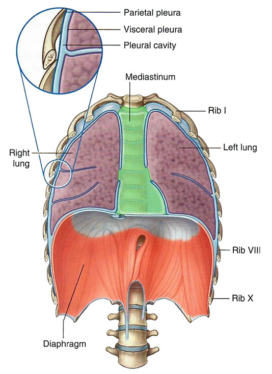
Although the pleura itself is not visible in the donor model, rotate the model to observe the potential spaces between the ribs and the lungs (costal surface) and the diaphragm and the lungs (diaphragmatic surface). These areas represent the pleural cavity, a potential space between the parietal and visceral pleura. Visualizing these relationships will help you understand the clinical significance of the pleural space.