Albany Medical College Virtual Anatomy Lab

SECTRA TABLE WORK: Page 2 of 6

The sinoatrial (SA) node is not visible in this donor model. Rotate the donor reconstruction and refer to the anatomical illustrations to identify its approximate location.
Add the atrioventricular (AV) node, highlighted in pink.

The AV node is located in the interatrial septum and is no longer visible once the septum is added.
Add the interatrial septum.
Add the interventricular septum.
conduction
Tap on image to enlarge
conduction
The AV (atrioventricular) node gives rise to the left and right bundle branches, which conduct impulses that coordinate contraction of the left and right ventricles. These branches are not visible in this individual.

Make certain to find some time to review the cardiac conduction system using the reconstructed models, plastinates, and anatomical illustrations.
Add the tricuspid valve.
Add the pulmonary valve.
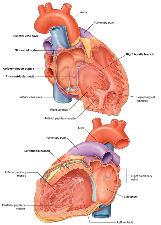
Add the pulmonary trunk and the left and right pulmonary arteries.
flow
Tap on image to enlarge
flow
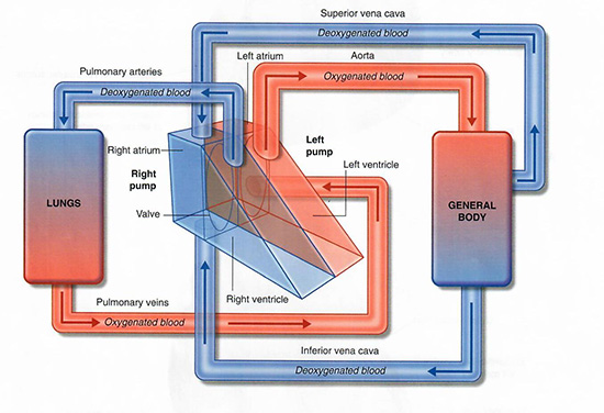
Briefly trace the path of blood flow from the superior and inferior vena cava through the tricuspid valve, into the right ventricle and out through the pulmonic valve into the pulmonary arteries.

Spend additional time exploring the flow of blood through the heart and associated vessels to solidify your understanding of this circulatory pathway.