
SECTRA TABLE WORK: Page 5 of 5
 |
| Tap on image to enlarge |
Let's examine the vascular supply:
| Rotate and remove the left thorax and the sternum to expose the right thorax. |
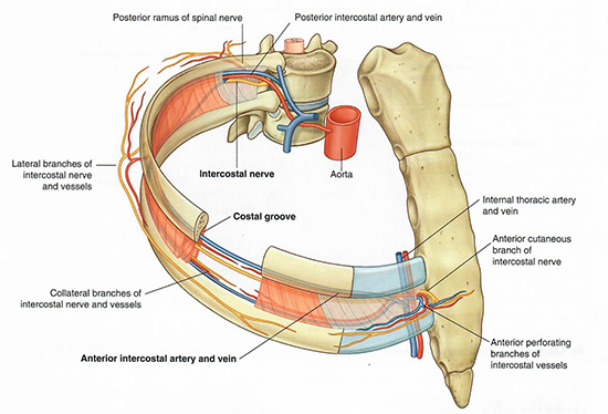
You will now add the components of the intercostal vessels and nerves.
Notice that the ventral ramus of the 1st thoracic spinal nerve (T1) runs across the superior surface of the first rib. To which major nerve network does it contribute? |
|
|
The brachial plexus. The T1 ventral ramus splits around the neck of the first rib. A major branch runs along the superior aspect of the first rib and contributes to the brachial plexus. In addition, a smaller branch becomes the 1st intercostal nerve and runs inferior to the first rib in the 1st intercostal space. |
|
Observe that each intercostal neurovascular bundle (vein, artery, and nerve) travels in the costal groove along the inferior border of the rib. Why is this clinically significant? |
|
|
The neurovascular bundle is vulnerable to injury during procedures such as thoracentesis or chest tube insertion. To avoid damage to these structures, clinicians are taught to insert instruments just above the superior border of the inferior rib. |
|
| Add the internal thoracic artery and vein, which run posterior (deep) to the costal cartilages and parallel to the sternum. |
| Re-add the sternum and costal cartilages. |
Rotate the chest to observe the anterior-to-posterior relationships of the thoracic wall structures.
| Add an axial cross-section. |
Now, rotate the 3D chest model slightly to bring the axial cross-section into view. In the side panel, examine the axial slice and identify thoracic wall structures you explored today, scrolling up and down the stack as needed.
Exploring the 3D model alongside its correlated cross-sections will strengthen your imaging interpretation skills. Nicely done!