Albany Medical College Virtual Anatomy Lab

Thorax Lab 1 Module 2
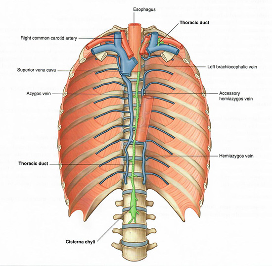
OBJECTIVES:

1.1.1   Identify the boundaries and divisions of the mediastinum and identify all structures in each part.

1.1.2   Demonstrate the projection of the thoracic diaphragm on the thoracic wall.

1.1.3   Follow the course and show the anatomical relationships of the phrenic nerve from its origin in the neck to the diaphragm.

1.1.4   Trace the course of the vagus nerve and its branches through the mediastinum.

1.1.5   Identify the recurrent laryngeal, cardiac, pulmonary, and esophageal branches of the vagus nerve and describe their target organs.

1.1.6   Identify the sympathetic trunk and the greater splanchnic nerve.

1.1.7   Trace the flow of blood in the azygos system and indicate any collateral connections to other parts of the systemic venous system.

1.1.8   Trace the flow of lymph through the thoracic duct from its origin to its termination, and indicate the sources of afferent lymphatics.

SECTRA TABLE WORK: Page 1 of 6

Azygous
Begin with the thoracic cage. AzygousTap on the image to enlarge.
Add the thoracic duct.
Observe that the thoracic duct begins in the abdomen at the cisterna chyli. Note that the term chyle refers to the milky white fluid produced in the small intestine during digestion, composed of lymph and emulsified fats. The thoracic duct ascends through the thorax and typically empties into the left venous angle, at the junction of the subclavian and left internal jugular veins (but sometimes into the brachiocephalic vein).
Add the azygos vein.
The name of this vein reflects its structure: zygos means paired; thus, azygos means unpaired. You can see the remnants of the original paired system in the hemiazygos vein. The azygos vein passes behind the root of the right lung before arching over it to empty into the superior vena cava (or SVC).
Add the diaphragm.
Notice that both the hemiazygos veins and the thoracic duct leave the abdomen near the crura of the diaphragm, muscular structures that also allow the aorta to pass from the thorax into the abdomen. Rotate the donor reconstruction to visualize this anatomical relationship more clearly.