Now, let's build the components of the mediastinum.
Start by adding the esophagus. Identify its course through the thorax, posterior to the trachea and heart.
Add the tracheobronchial tree. Visualize the branching of the trachea into the primary bronchi and their subdivisions within the lungs.
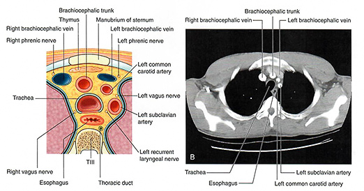
Add the major arteries of the thorax. Explore the aorta and its major branches: the brachiocephalic trunk, left common carotid artery, and left subclavian artery.
Overlay the axial cut represented by the blue line. This line corresponds approximately to the level of the images, below.
Compare the axial view of the VHD donor to the images. Use this comparison to better understand the anatomical structures visible at this level and how they appear on clinical imaging.