
Pelvis and Perineum: Module 1 - Page 2 of 5
Now, add the muscles and ligaments that form the lateral walls and define the apertures of the pelvic cavity.
| Rotate the 3D model to examine each item thoroughly, and identify the apertures of the pelvic wall. These include the greater sciatic foramen, the lesser sciatic foramen and the obturator canal. |
|
In the image below, observe the structures that pass through these apertures. 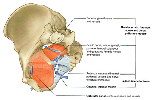 |
The lesser sciatic foramen is the primary entry for neurovascular structures entering the perineum (e.g. pudendal nerve and internal pudendal vessels). The greater sciatic foramen and obturator canal primarily allow communication between the pelvic cavity and the gluteal region of the lower limb.