
Male Pelvis and Genitalia: Page 5 of 8
| Add the patent portion of the umbilical artery. | |
| Add the obliterated (occluded) portion of the umbilical artery. |
The umbilical arteries are functional in fetal circulation, returning deoxygenated blood to the placenta. After birth, the distal portions obliterate and form the medial umbilical ligaments. These arteries will be removed during the next step for enhanced clarity.
| Add the spermatic cord. |
What structures are contained within the spermatic cord? |
|
|
The spermatic cord contains the: 1. ductus (vas) deferens 2. artery to the ductus deferens (or deferential artery) 3. testicular artery (from the abdominal aorta) 4. cremasteric artery and vein (from the inferior epigastrics) 5. genital branch of the genitofemoral nerve (innervates the cremaster muscle) 6. autonomic fibers (including sympathetics and visceral afferents) 7. Lymphatic vessels (draining to the lumbar, para-aortic, lymph nodes 8. Remnants of the processus vaginalis. Note that a patent processus vaginalis can lead to an indirect inguinal hernia or hydrocele. |
|
Note: Fibers of internal oblique muscle contribute to the cremaster muscle, which surrounds the spermatic cord and testes. The cremaster helps form part of the deep inguinal ring.
| Add the external abdominal oblique muscle. |
Note: The aponeurosis forms the inguinal ligament and superficial inguinal ring.
| Identify the inferior epigastric arteries and recall that they are a critical surgical and anatomical landmark. The deep inguinal ring lies just lateral to the inferior epigastric vessels. This relationship helps distinguish indirect inguinal hernias (lateral to the vessels) from direct inguinal hernias (medial to the vessels). Use the 3D model to visualize this anatomy and then compare it to the accompanying diagram of the inguinal canal and its contents. |
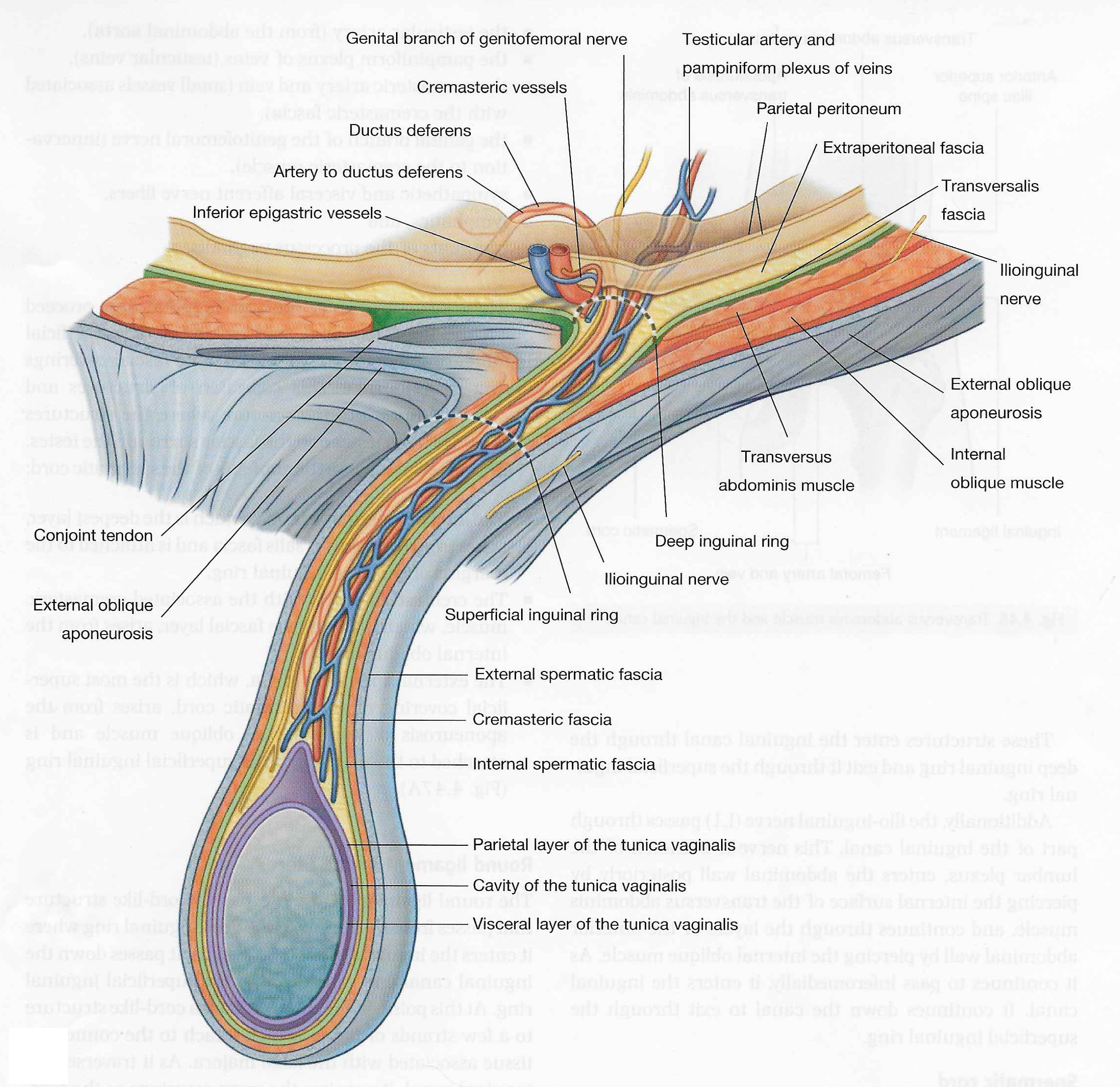 |
| Contents of Inguinal Canal Tap on image to enlarge |
/>