
Neurology Lab 1 - Module 2 - Ventricles of the Brain: Page 1 of 2
×
![Image]()
READINGS:Gray's Anatomy for Students (Fourth Edition): Pages: 865 |
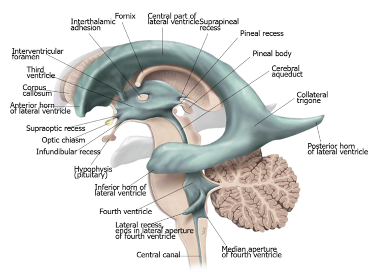 |
| Tap on image to enlarge |
| Begin with the Brain. |
| Remove the Left hemispheres of the cerebrum and cerebellum. |
| Highlight the lateral ventricles. |
| Highlight the third ventricle. |
| Highlight the fourth ventricle. |
| Remove the Right hemispheres of the cerebrum and cerebellum. |
| Remove the Vermis of the Cerebellum. |
| Remove the Medulla oblongata (Myelencephalon). |
| Remove the Pons. |
| Remove the Midbrain (Mesencephalon). |
| Remove the Left and Right Thalamus. |
| Remove the Anterior Commissure. |
| Remove the Corpus Callosum. |
| Please note: the ventricles are connected in life. However, the connections between the ventricles are not visible in this donor. Small channels such as the interventricular foramina and the cerebral aqueduct (of Sylvius) are not visualized due to their tiny size. |