Albany Medical College Virtual Anatomy Lab

Neurology Lab 1 - Module 1 - Blood Supply to the Brain: Page 4 of 5

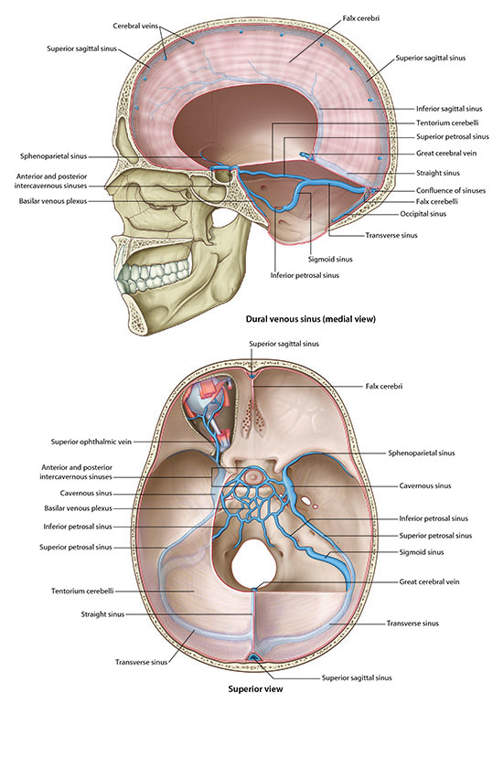
Next, examine the venous drainage of the brain.

Reflections of the meningeal layer of dura mater form the falx cerebri (within the longitudinal fissure) and the tentorium cerebelli (between the occipital lobes and cerebellum). These dural folds contain the major dural venous sinuses.

Dura venous sinus - medial view and superior view
Tap on image to enlarge
Dura venous sinus - medial view and superior view
Begin with the superior sagittal sinus, located along the attached superior margin of the falx cerebri.
Add the left and right transverse sinus, coursing laterally from the confluence along the attached margin of the tentorium cerebelli.
Add the confluence of the sinuses, near the internal occipital protuberance.
Add the straight sinus formed by the union of the inferior sagittal sinus and great cerebral vein. It courses posteriorly to drain into the confluence.
Add the inferior sinus in the free inferior margin of the falx cerebri. It empties into the straight sinus.
Add the sigmoid sinuses, which exit the skull via the jugular foramen and continue as the internal jugular veins.

Between what layers of the meninges are the dural venous sinuses located?