Lab 8 - Module 1 - Anatomy of the Knee: Page 8 of 10
×
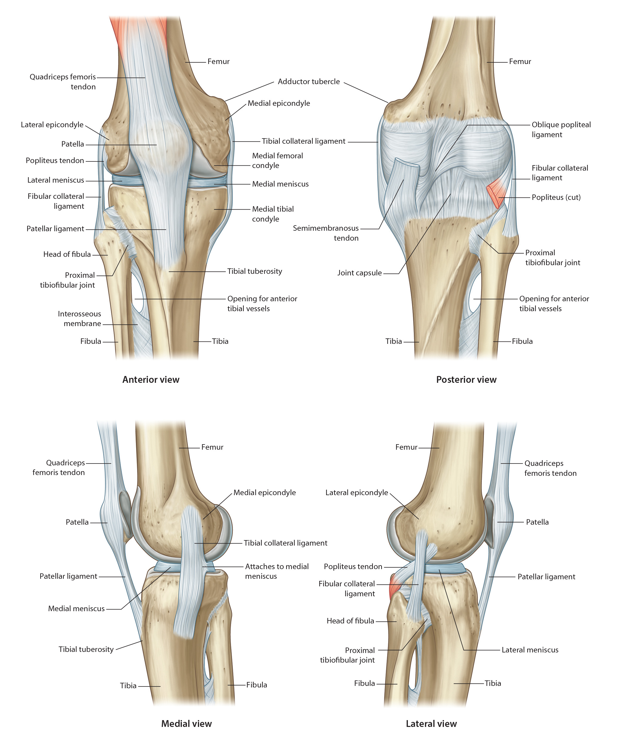
Ligamentous Support of the Knee Joint
Tap on image to enlarge
Add the Lateral Collateral Ligament (Fibular Collateral Ligament) which is distinct from the capsule and spans from the lateral epicondyle of the femur to the head of the fibula. It will resist adduction of the tibia on the femur.
Posterior reinforcements to the capsule
Add the Oblique Popliteal Ligament(NOT IDENTIFIED IN THIS CADAVER) which is said to be a continuation of the semimembranosis muscle, spanning downward with the tendon to insert on the medial aspect of the posterior side of the tibia, then continuing across the back of the joint to attach to the lateral femoral condyle on its posterior surface. Its function is to limit external rotation of the tibia on the femur and to limit knee extension and hyperextension.
Add the Arcuate ligament (NOT IDENTIFIED IN THIS CADAVER) which has two bands which both originate at the head of the fibula posteriorly. The lateral head spans directly upward to insert on the lateral aspect of the femur, while the medial head travels over the popliteus muscle to attach in the intercondylar area on the posterior aspect of the knee. It will help stabilize the posterior lateral corner of the knee in cases of rotary instability. The arcuate ligament provides a pathway for the popliteus muscle to enter the joint capsule.
Reinforcement of the Medial Aspect of the Capsule
Add the Medial Collateral Ligament (MCL) (Tibial Collateral Ligament) which has two layers.
The superficial layer spans from the medial epicondyle of the femur downward to the medial tibial flare.
The deep portion of the MCL originates at the same point and passes downward and posteriorly to insert along the medial tibial condyle. The deep layer is known as the Posterior Oblique Ligament.
The two layers are separated from each other by a bursa. Both portions will get taut with knee extension and both will help to limit abduction of the tibia on the femur.