Lab 8 - Module 1 - Anatomy of the Knee: Page 2 of 10
×
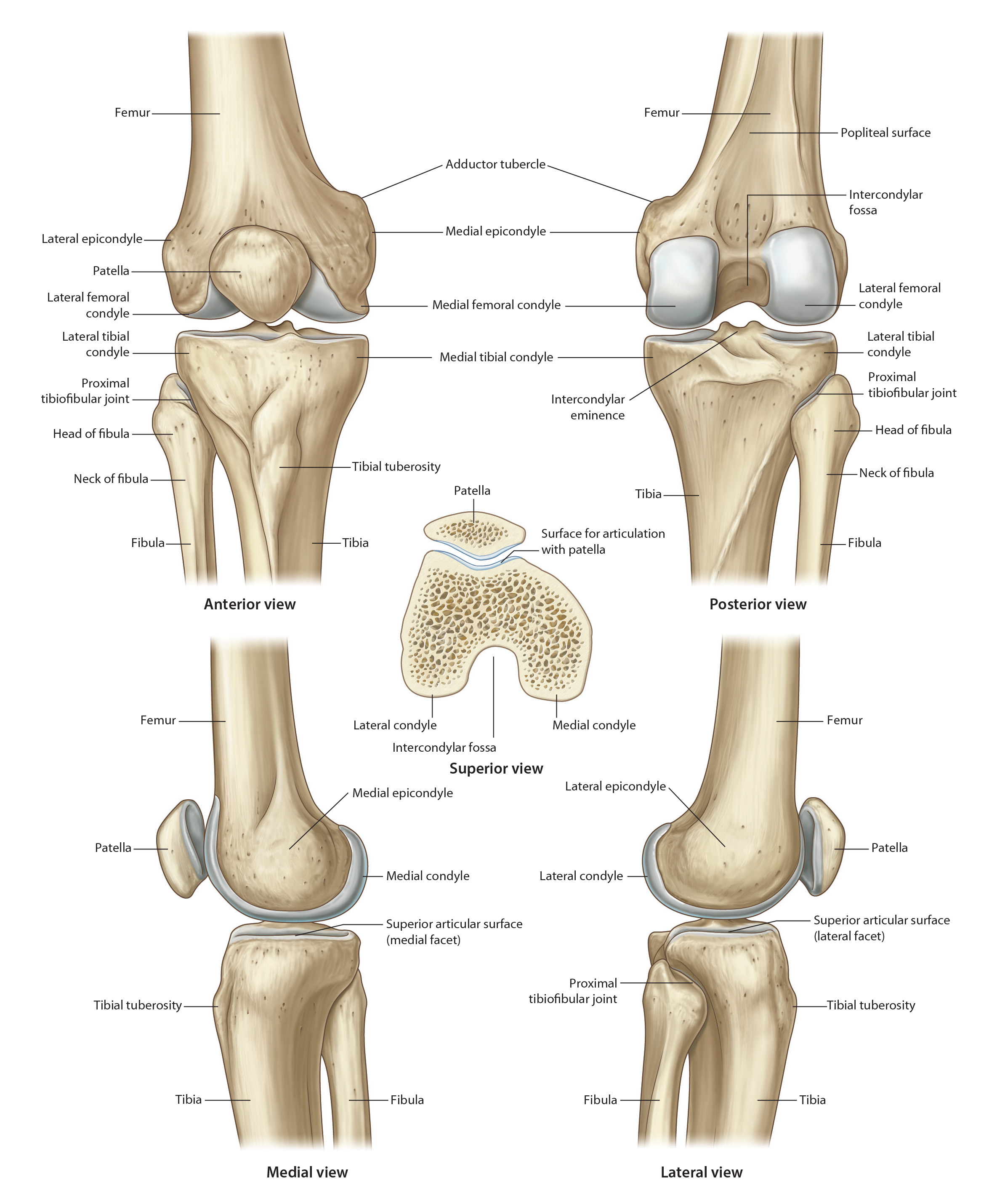
Osteology of the Knee
Tap on image to enlarge
There are four bones associated with the knee joint.
Begin with the femur. (Rotate the femur to visualize the following land marks:)
The lower end of the femur comprises two prominences, the lateral and medial femoral condyles. The femoral condyles are continuous superiorly with the femoral shaft. Each condyle is curved from front to back, and may be described as being cam-shaped.
Posteriorly, and to an extent inferiorly, they are separated from each other by a deep notch termed the intercondylar fossa (or intercondylar notch). The anterior, inferior and posterior aspects of each condyle are smooth and represent the articular surface. In life, this area is covered by articular hyaline cartilage. Viewed end-on, the articular surface on the distal (condylar) end of the femur is in the shape of a broad, inverted letter U. The articular surfaces of the medial and lateral femoral condyles engage, respectively, the meniscus-bearing medial and lateral articular facets on the tibial plateau.
The lateral surface of the lateral condyle and the medial surface of the medial condyle are non-articular, and both surfaces are readily palpable. The epiphyseal line at the lower end of femur is above the level of the femoral condyles.