Lab 5 - Module 1 - Hip and Anterior Thigh - Osteology of the Hip and Thigh: Page 4 of 6
×
Tap on image to enlarge
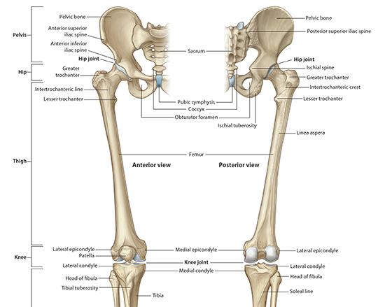
The Femur:
The femur which articulates with the acetabulum of the coxal bone to form the hip joint. The head of the femur fits into the acetabulum deeply. The femur corresponds to the humerus and the acetabulum corresponds to the glenoid fossa of the scapula.
Head - ball like that is covered with hyaline cartilage. It has a depression on the medial side (fovea capitis). The fovea serves as the attachment for the ligament of the head. The head fits into the cup like acetabulum to form the hip joint.
Neck- smaller in diameter than the head. Comes off at an angle to the long axis of the body which forms an angle of inclination (same as in the humerus). Head and neck may also be retroverted (facing posteriorly) or anteverted (facing anteriorly), these angles are referred to as torsion angles.
Greater Trochanter (and trochanteric fossa) - projection laterally off the junction of the neck and the body. Important site for muscle attachment. It also has a small depression on the medial aspect called the Trochanteric Fossa which serves as an important site for muscle attachment.
Lesser Trochanter - smaller projection off the junction of the head and neck that is more posterior and medial than the greater trochanter. Also, a site of muscle attachment.
Intertrochanteric Crest - posterior line that connects the two trochanters. It serves as the site of attachment for the posterior joint capsule of the joint capsule.
Intertrochanteric Line - anterior line (not as well developed as the crest) that connects the two trochanters anteriorly. It serves as the anterior attachment for the joint capsule.
Gluteal Tuberosity - found at about the same level as the lesser tuberosity but lies strictly posteriorly. At times it is well developed and referred to as the third trochanter. It serves as an attachment for gluteus maximus.
Body - cylindrical in shape and runs from below the trochanters inferior to the condyles. Linea Aspera is the ridge that runs the length of the body (posteriorly) that is important for muscle attachment.
Condyles - rounded ends that are at the distal most aspect of the femur. They are covered in hyline cartilage and articulate with the tibial plateau to form the knee joint. Between the two condyles (anterior) is the articular surface of the patella. Posterior the condyles are separated by the intercondylar fossa.
Epicondyles - roughened areas on each of the condyles (lie superior to the condyle). Superior to the medial epicondyle is the adductor tubercle. These areas are all important for muscle and ligament attachment.