Lab 5 - Module 1 - Hip and Anterior Thigh - Osteology of the Hip and Thigh: Page 2 of 6
×
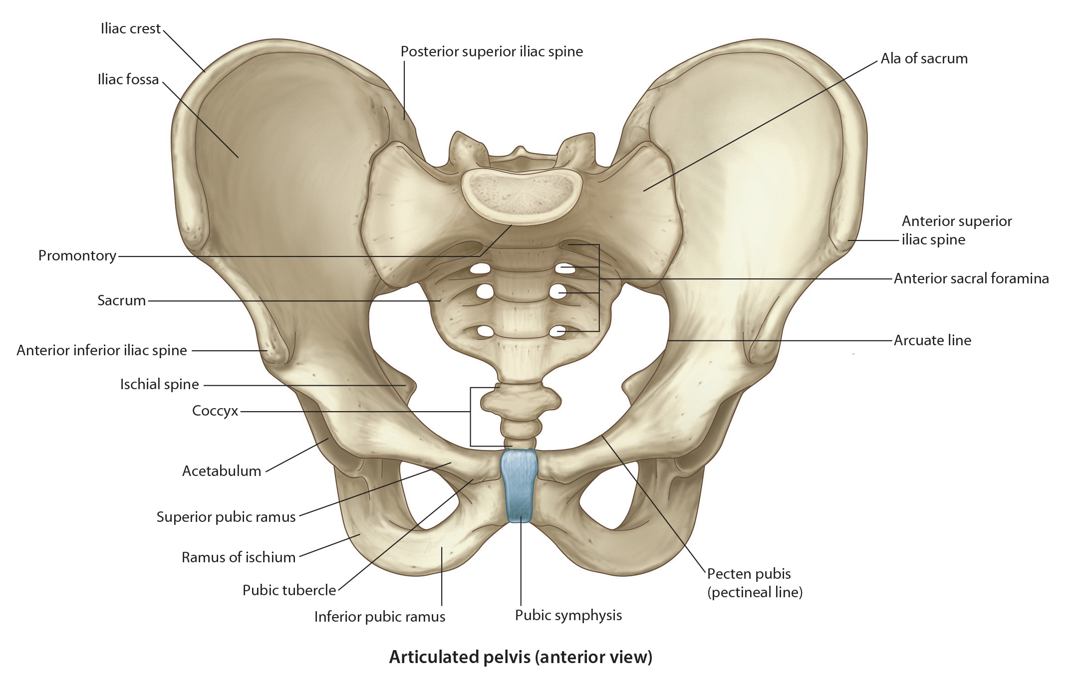
Osteology of the Hip
Tap on image to enlarge
Begin with the Pelvic Girdle.
The skeleton of the lower limb has similarities to that of the upper limb. The main difference is the need for the lower limb to weight bear. Weight bearing calls for increased stability when compared to the upper extremity. The main structural difference is that of the pelvic girdle.
The Pelvic Girdle is firmly attached to the vertebral column adding to the stability. The pelvic girdle is made up of three paired bones:
Ilium
Ishium
Pubis
These three bones are fused to form each of the coxal (hip) bones. The two coxal bones are firmly articulated to each other anteriorly and posteriorly and are firmly articulated with the sacrum.
The pelvis is capable of tilting (rotation) movements. Downward rotation is accompanied by an increase in the curvature of the lumbar spine (increased lordosis). Upward rotation decreases the lumbar curvature (decreased lordosis). The pelvis can also tilt laterally when body weight is supported on one limb.
Two functions of the pelvis:
1) transmit the weight of the body to the lower limbs
2) form the lower part of the abdominal cavity
The Coxal (Hip) Bones: formed by the fusion of the ilium, ishium, and the pubis. Ilium- it is the superior portion of the coxal bone and forms the superior ½ of the acetabulum. It forms the deep lateral aspect of the acetabulum. It consists of both a body and a wing. The body is the part that helps create the acetabulum and has no named parts. The wing consists of the: