Albany Medical College Virtual Anatomy Lab

Lab 4 - Module 2 - The Hand: Page 9 of 12

Interosseus and Lumbrical Muscles


Add the Dorsal Interossei Muscles. Each dorsal interossei (there are four of them) originates from two heads one from each of the adjacent metacarpal bones. Example: 1st dorsal interosseous muscle arises from the first and second metacarpal bones. The 1st dorsal interosseous muscle inserts into the extensor expansion on the proximal phalanx of the 2nd digit. The 2nd and 3rd interossei insert on the medial and lateral aspect of the proximal phalanx of the middle finger. The 4th interosseous inserts on the ulnar side of the ring finger. The deep branch of the ulnar nerve (C8, T1) innervates all four of the dorsal interossei muscles. Each abducts the fingers using the middle finger as the midline. It also helps to flex the metacarpophalangeal joints and extend the interphalangeal joints.

We can remember the actions of the palmar and dorsal interossei muscles through the memory of P.A.D and D.A.B.

P.A.D – Palmar Interossei ADduct
D.A.B – Dorsal Inersossei ABduct


Add the Palmar Interosseous Muscles.

First palmar interosseous muscle- it originates from the ulnar and palmar surfaces of the second metacarpal. It inserts on the ulnar aspect of the extensor expansion of the second digit. It is innervated by the the deep branch of the ulnar nerve (C8, T1). The first palmar interosseous adducts the index finger at the MP joint.

Second palmar interosseous muscle- it originates on the radial side of the fourth metacarpal. It inserts on the radial aspect of the extensor expansion of the fourth digit. It is innervated by the deep branch of the ulnar nerve. The second palmar interosseous adducts the ring finger at the MP joint.

Third palmar interosseous muscle- it originates on the radial side of the fifth metacarpal. It inserts on the radial aspect of the extensor apparatus of the fifth digit. It is innervated by the deep branch of the ulnar nerve. The third palmar interosseous adducts the little finger at the MP joint.

The three palmar interossei adduct the fingers relative to the midline of the long finger. Through their attachments to the extensor expansion, they contribute to flexion of the MP and extension of PIP and DIP joints. They also have a role in isolated rotation of a digit.


Add the Lumbrical Muscles (Four of them): All of them originate from the tendon of the flexor digitorum profundus (they have the same innervations as flexor digitorum profundus). They insert on the radial side of the 2nd, 3rd, 4th, and 5th extensor expansion, respectively. Each lumbrical is named for the digit it inserts into (E.G. the first lumbrical inserts on the second digit all the way to the fourth lumbrical inserts on the fifth digit. The lateral two lumbricals are innervated by the median nerve and the medial two are innervated by the deep branch of the ulnar nerve. They flex the metacarpophalngeal joints and help extend the interphalangeal joints.
Add the Flexor Digitorum Profundus.
Add the Flexor Digitorum Superficialis.
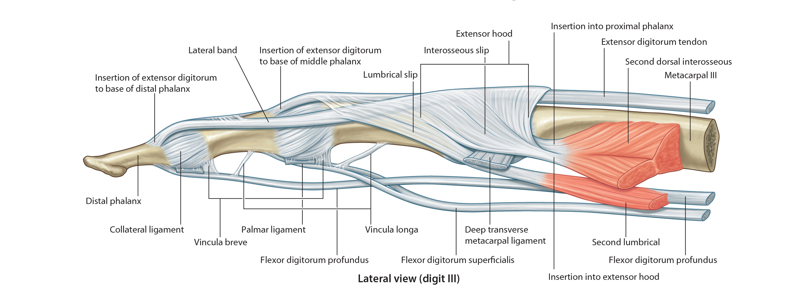
The tendons of these muscles pass in a common sheath deep to the flexor retinaculum. They then pass deep to the palmar aponeurosis and enter the osseofibrous tunnels digital tunnels. The palmar aponeurosis is the strong, well defined triangular part of the deep fascia covering the soft tissue of the hand. It overlies the long flexor tendons of the palm. The proximal end of the palmar aponeurosis is continuous with the flexor retinaculum.
Observe that there are two tendons in each osseofibrous tunnel. In order that these tendons can slide freely over each other during movements of the fingers, each tendon is covered with a synovial membrane. Near the base of the proximal phalanx, the tendon of the flexor digitorum superficialis splits and surrounds the tendon of the flexor digitorum profundus. The halves of the tendon of the flexor digitorum superficialis insert into the margins of the middle phalanx. The tendon of the flexor digitorum profundus, after passing through the split in the tendon of the flexor digitorum superficialis, passes distally to insert into the base of the distal phalanx.

With this patient, can you explain why the corticosteroids provided relief for a period of time, but the symptoms returned?

profunda arterial supply