Identify Distal Radioulnar Joint – a pivot type of synovial joint the proximal radioulnar joint. It involves the head of the ulna articulating with the ulnar notch in the distal end of the radius. A fibrocartilaginous articular disc binds the ends of the ulna and radius together and is the main uniting structure of the joint. The synovial capsule lines the fibrous capsule.
Pronation and supination occur at the distal radioulnar joint as they do at the proximal radioulnar joint. It is the radius that rotates over the ulna.
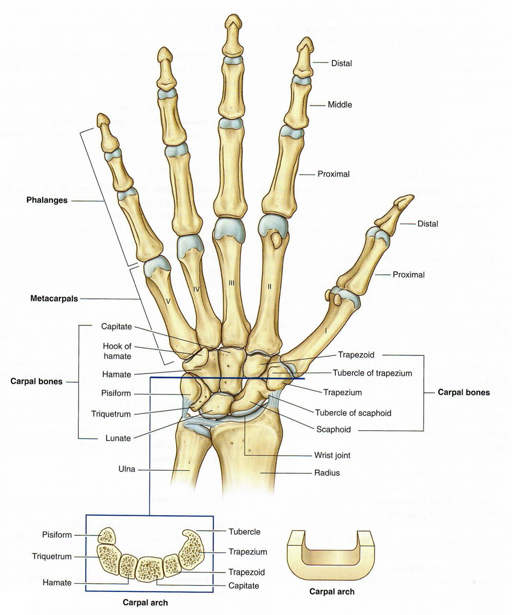
Identify the Wrist Joint. The wrist or radiocarpal joint is between the distal end of the radius and the proximal row of the carpus. It is a condyloid or ellipsoid type of synovial joint.
The following movements are possible – adduction (ulnar deviation), abduction (radial deviation), flexion, extension, and circumduction.
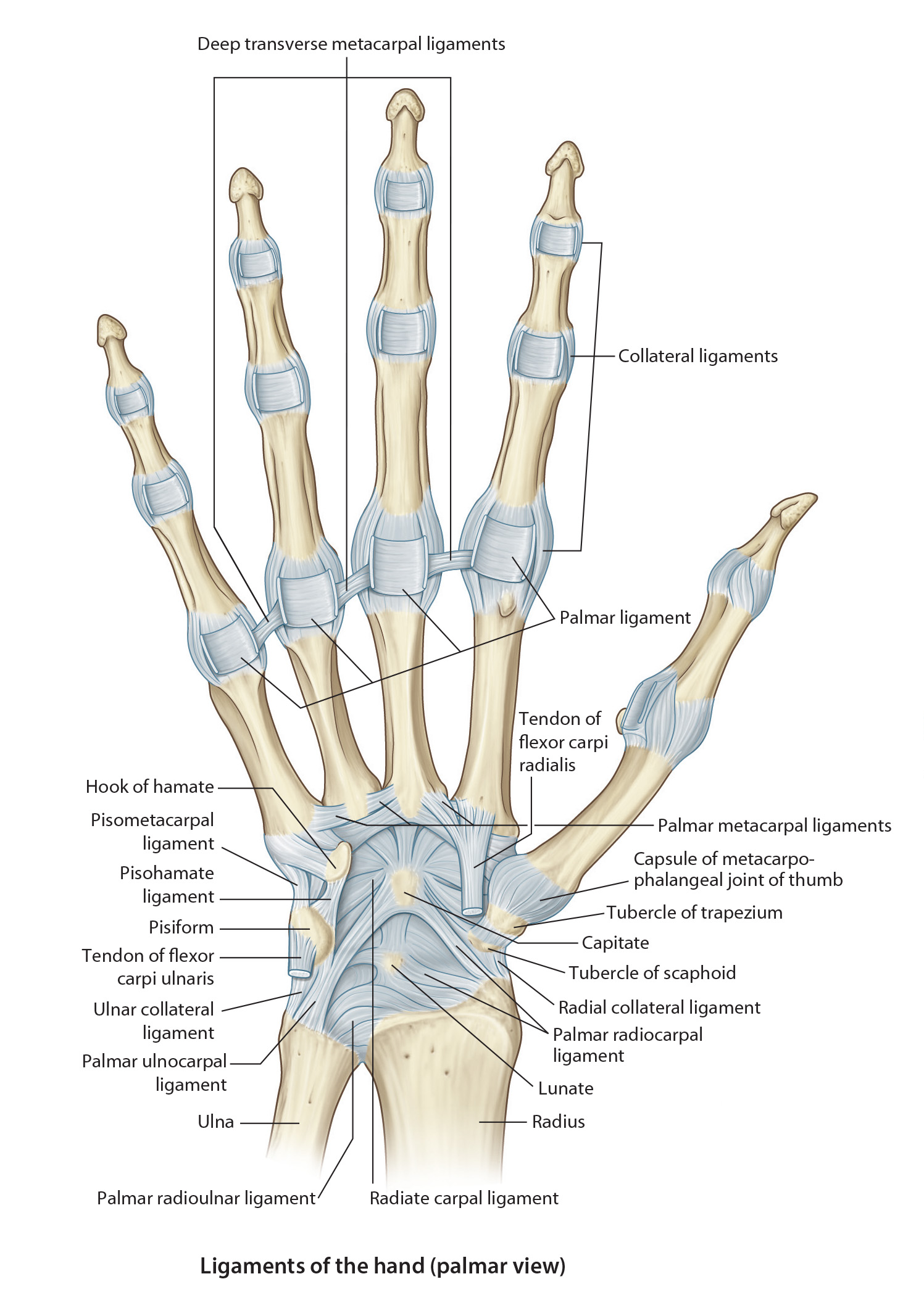
A fibrous capsule encloses the joint. It is strengthened by the dorsal and palmar radiocarpal ligaments. The fibrous capsule is also strengthened by the radial and ulnar collateral ligaments. The synovial capsule lines the fibrous capsule.
The blood supply is from the dorsal and palmar carpal arches.
The articular nerves are derived from the anterior interosseous branch of the median nerve, the posterior interosseous branch of the radial nerve and the dorsal and deep branches of the ulnar nerve.
Identify the Carpalpometacarpal Joints are plane synovial joints between the distal row of carpal bones and the bases of the metacarpal bones. These joints permit a small amount of gliding movement.
Carpometacarpal Joint of the Thumb is a separate saddle type synovial joint which permits angular movements in any plane and a restricted amount of axial rotation. The blood supply for the carpometacarpal joint of the thumb (AKA 1st carpometacarpal joint) is from the dorsal and palmar metacarpal arteries and from the dorsal carpal and deep palmar arches. Nerve supply is the same as the nerve supply for the wrist joint.
Identify the Metacarpophalangeal Joints – these articulations are the condyloid (knuckle-like) type of synovial joint that allow movement in two directions. The heads of the metacarpals articulate with the bases of the proximal phalanges.
The following movements occur at the metacarpophalangeal joints (MCP Joints): flexion, extension, abduction, adduction.
A fibrous capsule encloses each joint. They are strengthened on each side by a triangular collateral ligament. The palmar ligaments are strong thick plates that are firmly attached to the phalanx and loosely attached to the metacarpal. The palmar ligaments of the second to fifth joints are united by deep transverse metacarpal ligaments which hold the heads of the metacarpals together.
The articular arteries are branches of the digital arteries arising from the superficial palmar arch.
The articular nerves are derived from the digital nerves which arise from the ulnar and medial nerves.
Identify the Interphalangeal Joints – these articulations are uniaxial hinge joints which permit only flexion and extensions. There are proximal interphalangeal joints that are the articulation of the heads of the proximal phalanges and the bases of the middle phalanges of the medial four fingers. The thumb only has an interphalangeal joint because it only has two phalanges. There are distal interphalangeal joints that are the articulation of the heads of the middle phalanges and the bases of the distal phalanges of the medial four fingers. They are structurally similar to the MCP’s.
The articular arteries are branches of the digital arteries arising from the superficial palmar arch.
The articular nerves are derived from the digital nerves which arise from the ulnar and medial nerves.