Lab3 - Module 2 - Anatomy of the Arm and Forearm: Page 1 of 11
READINGS:
Gray's Anatomy for Students (Fourth Edition): Pages: 753-775, 782-793
The Flexor Forearm
Tap on image to enlarge
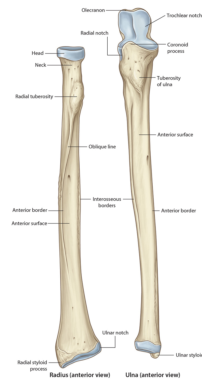
The forearm extends from the elbow to the wrist and contains two bones, the radius and ulna, which are parallel in the anatomic position. In pronation the radius lies across the ulna. The radius is the moveable bone of the forearm while the ulna remains stationary.
Begin with the bones of the forearm. The ulna is more firmly connected to the arm bone or humerus, whereas the radius is broadened distally to be more fully in contact with wrist bones. (It may help to think of the radius and hand as a unit when considering muscle actions.) Also note that the head of the ulna is at its distal end, whereas the head of the radius is at its proximal end.
Identify the Radius. The proximal end of the radius has a disc-shaped head, a smooth neck, and a tuberosity distal to the neck. The body (shaft) of the radius increases in size from proximal to distal. The medial aspect of the body has a sharp interosseous border for attachment of the interosseous membrane. The distal end has a median ulnar notch into which the distal end of the ulna fits. Laterally, the distal end of the radius tapers into a styloid process.
Identify the Ulna. The ulna is the longer and medially located bone of the forearm. The proximal end of the bone has the olecranon process, coronoid process and the trochlear notch between them. At the distal end lies the head. The lateral side of the coronoid process has a small radial notch for the head of the radius. Inferior to the radial notch is the supinator fossa, bounded posteriorly by a supinator crest. The anterior surface of the coronoid process is rough and ends distally in the tuberosity of the ulna onto which the brachialis muscle inserts. The body of the ulna has a prominent lateral edge, the interosseous border, where the interosseous membrane attaches. The distal end of the ulna is composed of a head and a styloid process, which projects distally about 1 cm proximal to the styloid process of the radius (this is clinically important).