Albany Medical College Virtual Anatomy Lab

Lab 1 Module 3: Posterior Triangle of the Neck - Page 4 of 5

Nerves of the Posterior Triangle


cervicalplexus
cervicalplexus
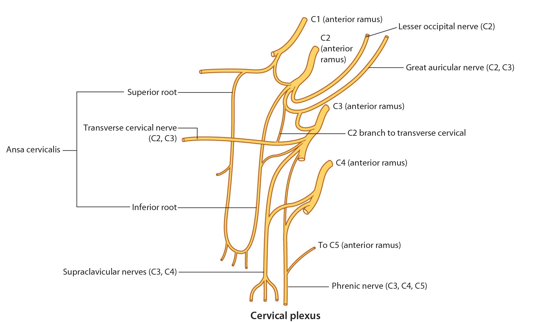
Tap on image to enlarge
The Cervical Plexus is a network of nerves formed by communications between the ventral rami of the first four cervical nerves (C1-C4). It has a sensory portion and a motor portion. The motor portion innervates muscles in the anterior triangle of the neck. The sensory portion emerges from deep to sternocleidomastoid, pierces the deep fascia and divides into four main sensory nerves:
The Lesser Occipital Nerve (C2) – ascends a short distance along the posterior border of the sternocleidomastoid muscle before dividing into several branches that supply the skin of the neck and scalp. (This nerve is not visable on this cadaver).
The Greater Auricular Nerve (C2-C3) – runs vertically over the sternocleidomastoid muscle toward the parotid gland (which lies anterior to the ear). It supplies branches to the skin of the neck, posterior aspect of the auricle of the ear, and an area extending from the mandible to the mastoid process. (This nerve is not visable on this cadaver).
The Transverse Cervical Nerve (C2-C3) – runs transversely across the middle of the sternocleidomastoid muscle to supply skin over the anterior triangle of the neck. (This nerve is not visable on this cadaver).
The Supraclavicular Nerves (C3-C4) – arise as a single trunk and divides into three main branches. They supply skin over the anterior aspect of the chest and shoulder.
The Phrenic Nerve (C3, C4, C5) – NOT part of the cervical plexus but derived from the ventral rami of C3, C4, C5. It runs on the anterior aspect of the anterior scalene muscle to enter the thoracic cavity where it ultimately innervates the diaphragm muscle (Mnemonic: C3, C4, C5 keeps the diaphragm alive).