
Lab 1 Module 3: Posterior Triangle of the Neck - Page 1 of 5
READINGS:Gray's Anatomy for Students (Fourth Edition): Pages: 1012-1019 |
The neck is divided into several triangles for descriptive purposes. The two main divisions are the Anterior Triangle and the Posterior Triangle. Each of these triangles can be further subdivided into smaller sub-triangles. Let's first focus on the Posterior Triangle.
 |
| Tap on image to enlarge |
|
Origin - manubrium of the sternum and medial 1/3rd of the clavicle. Insertion - mastoid process of the temporal bone the superior nuchal line of the occiput. Innervation - (Spinal) Accessory Nerve (Cranial Nerve XI), with proprioceptive fibers from ventral rami of C2-C3. Action - Unilaterally causes the contralateral rotation of the face and ipsilateral neck side bending. When acting bilaterally they help flex the neck and elevate the sternum during forced inspiration. |
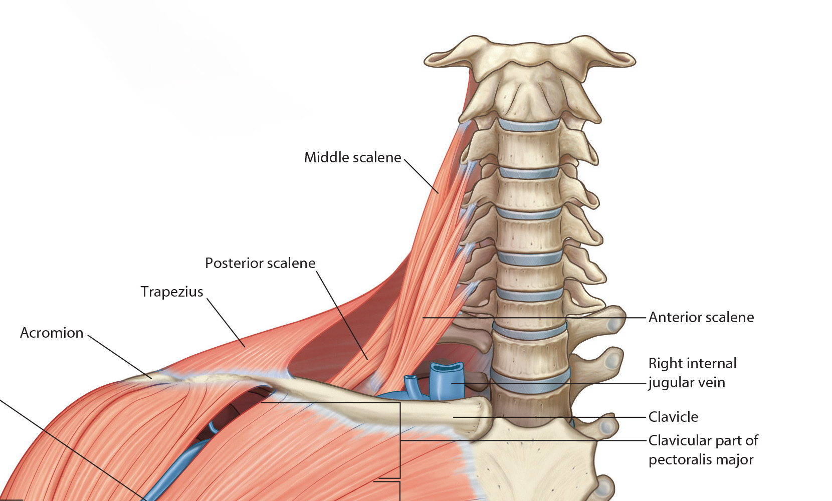
| Add the Posterior Scalene. |
|
Origin - Posterior tubercles of transverse processes of C5-C7 vertebrae. Insertion - external border of the second rib. Innervation - ventral rami of C7 and C8 cervical nerves. Action - laterally flexes the neck; elevates the second rib during forced inspiration. |
| Add the Middle Scalene. |
|
Origin - Posterior tubercles of transverse processes of C5-C7 vertebrae. Insertion - superior surface of the first rib, posterior to the groove for subclavian artery. Innervation - ventral rami of cervical spinal nerves (C3-C8). Action - flexes the neck laterally; elevates the first rib during forced inspiration. |
| Add the Anterior Scalene. |
|
Origin - Transverse processes of C3-C6 Insertion - Scalene tubercle of 1st Rib Innervation - ventral rami of C4-C6 Action - Flexes the head and assists with elevation of first rib during inspiration. |