
Lab 1 - Module 1: Pectoral Region - Page 1 of 5
READINGS:Gray's Anatomy for Students (Fourth Edition): Pages: 133-144 |
CASE REPORT:A 51 y/o woman presents to the office with a 1-month history of a lump in her right breast which she discovered during breast self-examination. The lump is not tender and has not changed in size since she first noticed it. She reports no skin changes or nipple discharge. She has been in good health and has no significant medical history. She had her first menstrual period at age 13 and has had three full-term pregnancies, the first at age 22. She reached menopause 3 years ago. She has no family history of breast or ovarian cancer and has never had a mammogram. Her last gynecological examination was 1 year ago and was normal. She takes no medications or hormonal supplements. Her part medical and surgical histories are noncontributory. |
EXAMINATION: |
Inspection while seated: |
Inspection while supine: |
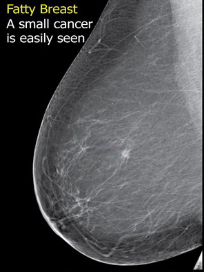 |
| Tap on image to enlarge |
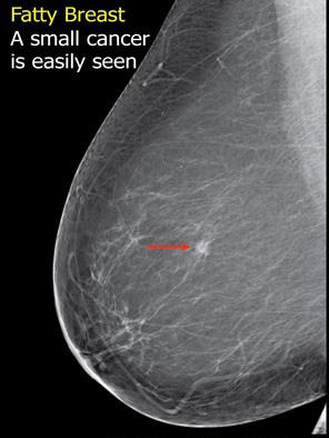
| A Mammogram was ordered confirming the mass. |
Can you identify the mass on the film? |
|
|
|
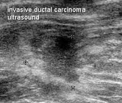 |
| Tap on image to enlarge |
| An Ultrasound Examination was ordered to determine if the mass was solid or a fluid-filled cyst. It was confirmed to be a solid mass. |
Can you identify the mass on the ultrasound? |
|
|
The stars on the scan point to the boundaries of the mass and the infiltration of the tumor into the surrounding tissue. |
|
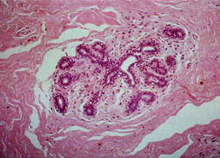 |
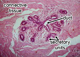
| Tap on image to enlarge |
| A Biopsy of a nromal breast looks like this slide. |
Can you identify the components of a normal Mammary gland on this slide? |
|
|
|
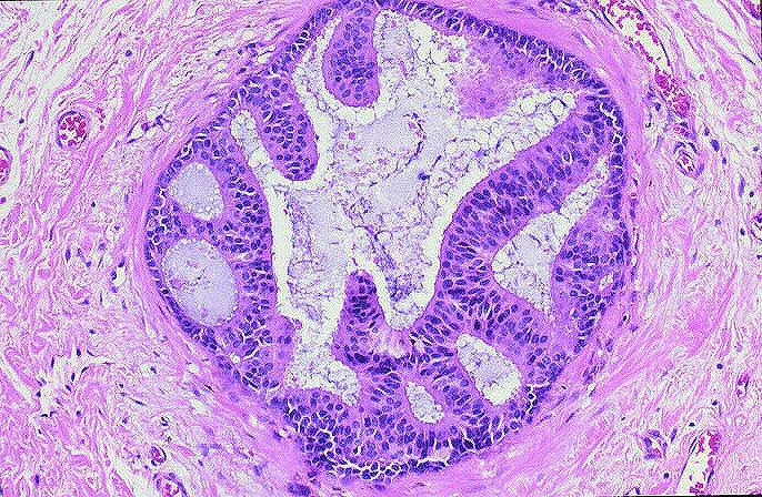
What does a invasive ductal carcinoma look like on a biopsy slide? |
|
|
|