
Thorax Lab 2 Module 2
OBJECTIVES:1.3.1 Trace the flow of blood through the major coronary arteries, identifying the origin, course, and distribution of each vessel. 1.3.2 Describe the venous drainage of the heart including the location and course of the major cardiac veins, and explain their relationship to the coronary arteries and their drainage into the coronary sinus. 1.3.3 Identify and distinguish the fibrous, parietal, and visceral layers of the pericardium, and describe their anatomical relationships and functional roles. |
SECTRA TABLE WORK: Page 1 of 4
| Begin with the heart. | |
| Add the major vessels entering and leaving the heart. |
 |
| Tap on image to enlarge |
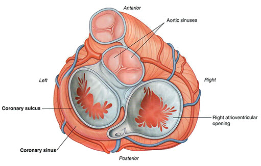
How may coronary artery openings (ostia) are there in the aortic trunk? |
|
|
There are two coronary ostia, one for the right coronary artery and one for the left coronary artery. |
|
Where are these openings located in relation to the aortic valve? |
|
|
The coronary ostia are located in the aortic sinuses, just proximal (with regard to blood flow) to the cusps of the aortic valve. Specifically, each ostium lies just superior to and outside the corresponding aortic valve cusps, within the wall of the ascending aorta. 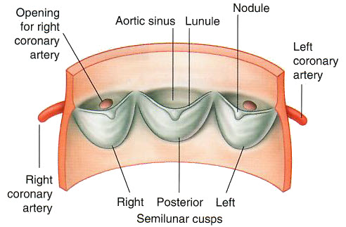
|
|