Albany Medical College Virtual Anatomy Lab

SECTRA TABLE WORK: Page 4 of 6

Rotate the donor reconstruction to visualize the azygos vein draining directly into the superior vena cava (SVC) just above the right bronchus.
Add the pulmonary veins. Observe how they return oxygenated blood from the lungs to the left atrium of the heart.
Add the lungs. Appreciate the relationship of the lungs with the surrounding structures.
Add the thymus, located just posterior to the sternum.
Review the boundaries of the mediastinum, which separates the two pleural cavities containing the lungs. It extends from the thoracic inlet to the diaphragm, and from the sternum to the thoracic vertebrae.

Rotate the model to explore the mediastinum and its contents.
Mediastinum
Tap on the image to enlarge
Mediastinum
Add the sternum and remove the left side of the thorax. This will expose the compartments of the mediastinum.
Using the Highlight and Draw tools, identify thoracic vertebrae T4 and T5. Then draw a line between the sternal angle and the T4/T5 intervertebral space. Everything superior to this line is in the superior mediastinum.
Using the Highlight and Draw tools, identify the anterior, middle and posterior compartments of the inferior mediastinum.

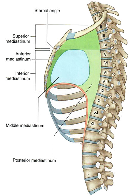
What structures are contained in the middle compartment of the mediastinum?

Through which mediastinal compartments does the esophagus pass?